Muscles of the Upper Limb - Listed Alphabetically
|
| Muscle |
Origin |
Insertion |
Action |
Innervation |
Artery |
Notes |
Image |
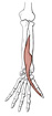
| abductor digiti minimi (hand) |
pisiform |
base of the proximal phalanx of the 5th digit on its ulnar side |
abducts the 5th digit |
deep branch of the ulnar nerve |
ulnar a. |
abductor digiti minimi, flexor digiti minimi brevis, and opponens digiti
minimi are located in the hypothenar compartment of the hand |
 |
| abductor pollicis brevis |
flexor retinaculum, scaphoid, trapezium |
base of the proximal phalanx of the first digit |
abducts thumb |
recurrent branch of median nerve |
superficial palmar br. of the radial a. |
abductor pollicis brevis, flexor pollicis brevis, and opponens pollicis
are located in the thenar compartment of the hand |
 |
| abductor pollicis longus |
middle one-third of the posterior surface of the radius, interosseous membrane,
mid-portion of posterolateral ulna |
radial side of the base of the first metacarpal |
abducts the thumb at carpometacarpal joint |
radial nerve, deep branch |
posterior interosseous a. |
the tendons of abductor pollicis longus and extensor pollicis brevis make
the lateral border of the anatomical snuffbox |
 |
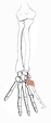
| adductor pollicis |
oblique head: capitate and base of the 2nd and 3rd metacarpals; transverse
head: shaft of the 3rd metacarpal |
base of the proximal phalanx of the thumb |
adducts the thumb |
ulnar nerve, deep branch |
deep palmar arterial arch |
deep palmar arch and deep ulnar nerve pass between the two heads of adductor
pollicis, which is in the adductor-interosseous compartment |
 |
| anconeus |
lateral epicondyle of the humerus |
lateral side of the olecranon and the upper one-fourth of the ulna |
extends the forearm |
nerve to anconeus, from the radial nerve |
interosseous recurrent a. |
none |
 |
| biceps brachii |
short head: tip of the coracoid process of the scapula; long head: supraglenoid
tubercle of the scapula |
tuberosity of the radius |
flexes the forearm, flexes arm (long head), supinates |
musculocutaneous nerve (C5,6) |
brachial a. |
a powerful supinator only if the elbow is flexed |
 |
| brachialis |
anterior surface of the lower one-half of the humerus and the associated
intermuscular septa |
coronoid process of the ulna |
flexes the forearm |
musculocutaneous nerve (C5,6) |
brachial a., radial recurrent a. |
a powerful flexor |
 |
| brachioradialis |
upper two-thirds of the lateral supracondylar ridge of the humerus |
lateral side of the base of the styloid process of the radius |
flexes the elbow, assists in pronation & supination |
radial nerve |
radial recurrent a. |
although brachioradialis is innervated by the nerve for extensors (radial),
its primary action is elbow flexion; the neutral position of this muscle is
half way between supination and pronation (elbow flexed, thumb up) |
 |
| coracobrachialis |
coracoid process of the scapula |
medial side of the humerus at mid-shaft |
flexes and adducts the arm |
musculocutaneous nerve (C5,6) |
brachial a. |
the musculocutaneous nerve passes through the coracobrachialis muscle to
reach the other arm flexor mm.(biceps brachii and brachialis) |
 |
| deltoid |
lateral one-third of the clavicle, acromion, the lower lip of the crest
of the spine of the scapula |
deltoid tuberosity of the humerus |
abducts arm; anterior fibers flex & medially rotate the arm; posterior fibers
extend & laterally rotate the arm |
axillary nerve (C5,6) from the posterior cord of the brachial plexus |
posterior circumflex humeral a. |
the deltoid muscle is the principle abductor of the arm but due to poor
mechanical advantage it cannot initiate this action; it is assisted by the
supraspinatus m. |
 |
| dorsal interosseous (hand) |
four muscles, each arising from two adjacent metacarpal shafts |
base of the proximal phalanx and the extensor expansion on lateral side
of the 2nd digit, lateral & medial sides of the 3rd digit, and medial side
of the 4th digit |
flex the metacarpophalangeal joint, extend the proximal and distal interphalangeal
joints of digits 2-4, abduct digits 2-4 (abduction of digits in the hand is
defined as movement away from the midline of the 3rd digit) |
ulnar nerve, deep branch |
dorsal and palmar metacarpal aa. |
bipennate muscles; remember DAB & PAD - Dorsal interosseous mm. ABduct and
Palmar interosseous mm. ADduct - then you can figure out where they must insert
to cause these actions |
 |
| extensor carpi radialis brevis |
common extensor tendon (lateral epicondyle of humerus) |
dorsum of the third metacarpal bone (base) |
extends the wrist; abducts the hand |
deep radial nerve |
radial a. |
works with the extensor carpi radialis longus and flexor carpi radialis in abduction of the hand |
 |
| extensor carpi radialis longus |
lower one-third of the lateral supracondylar ridge of the humerus |
dorsum of the second metacarpal bone (base) |
extends the wrist; abducts the hand |
radial nerve |
radial a. |
works with the extensor carpi radialis brevis and flexor carpi radialis
in abduction of the hand |
 |
| extensor carpi ulnaris |
common extensor tendon & the middle one-half of the posterior border of
the ulna |
medial side of the base of the 5th metacarpal |
extends the wrist; adducts the hand |
deep radial nerve |
ulnar a. |
works with the flexor carpi ulnaris in adduction of the hand |
 |
| extensor digiti minimi |
common extensor tendon (lateral epicondyle of the humerus) |
joins the extensor digitorum tendon to the 5th digit and inserts into the
extensor expansion |
extends the metacarpophalangeal, proximal interphalangeal and distal interphalangeal
joints of the 5th digit |
deep radial nerve |
interosseous recurrent a. |
extensor digiti minimi appears to be the ulnar-most portion of extensor
digitorum |
 |
| extensor digitorum |
common extensor tendon (lateral epicondyle of the humerus) |
extensor expansion of digits 2-5 |
extends the metacarpophalangeal, proximal interphalangeal and distal interphalangeal
joints of the 2nd-5th digits; extends wrist |
deep radial nerve |
interosseous recurrent a. and posterior interosseous a. |
the extensor expansion inserts via a central band on the base of the middle
phalanx, while lateral & medial slips insert on the distal phalanx |
 |
| extensor indicis |
interosseous membrane and the posterolateral surface of the distal ulna |
its tendon joins the tendon of the extensor digitorum to the second digit;
both tendons insert into the extensor expansion |
extends the index finger at the metacarpophalangeal, proximal interphalangeal
and distal interphalangeal joints |
deep radial nerve |
posterior interosseous a |
extensor indicis is a deep forearm extensor, whereas extensor digiti minimi
is in the superficial layer of extensors |
 |
| extensor pollicis brevis |
interosseous membrane and the posterior surface of the distal radius |
base of the proximal phalanx of the thumb |
extends the thumb at the metacarpophalangeal joint |
deep radial nerve |
posterior interosseous a |
the tendons of extensor pollicis brevis and abductor pollicis longus make
the lateral border of the anatomical snuffbox, in which the radial arterial
pulse can be felt |
 |
| extensor pollicis longus |
interosseous membrane and middle part of the posterolateral surface of the
ulna |
base of the distal phalanx of the thumb |
extends the thumb at the interphalangeal joint |
deep radial nerve |
posterior interosseous a |
the tendon of extensor pollicis longus hooks around the dorsal radial tubercle;
it forms the medial border of the anatomical snuffbox, in which the radial
arterial pulse can be felt |
 |
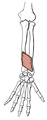
| flexor carpi radialis |
common flexor tendon from the medial epicondyle of the humerus |
base of the second and third metacarpals |
flexes the wrist, abducts the hand |
median nerve |
ulnar a. |
works with the extensor carpi radialis longus and brevis mm. to abduct hand |
 |
| flexor carpi ulnaris |
common flexor tendon & (ulnar head) from medial border of olecranon & upper
2/3 of the posterior border of the ulna |
pisiform, hook of hamate, and base of 5th metacarpal |
flexes wrist, adducts hand |
ulnar nerve |
ulnar a. |
the ulnar nerve passes between the two heads of origin of the flexor carpi
ulnaris m. |
 |
| flexor digiti minimi brevis (hand) |
hook of hamate & the flexor retinaculum |
proximal phalanx of the 5th digit |
flexes the carpometacarpal and metacarpophalangeal joints of the 5th digit |
ulnar nerve, deep branch |
ulnar a. |
flexor digiti minimi brevis, abductor digiti minimi, and opponens digiti
minimi are in the hypothenar compartment of the hand |
 |
| flexor digitorum profundus |
posterior border of the ulna, proximal two-thirds of medial border of ulna,
interosseous membrane |
base of the distal phalanx of digits 2-5 |
flexes the metacarpophalangeal, proximal interphalangeal and distal interphalangeal
joints |
median nerve (radial one-half); ulnar nerve (ulnar one-half) |
ulnar a., anterior interosseous a. |
ulnar nerve innervates the portion of profundus that acts on digits 4 &
5 (the ulnar 2 digits) |
 |
| flexor digitorum superficialis |
humeroulnar head: common flexor tendon; radial head: middle 1/3 of radius |
shafts of the middle phalanges of digits 2-5 |
flexes the metacarpophalangeal and proximal interphalangeal joints |
median nerve |
ulnar a. |
median nerve travels distally in the forearm on the deep surface of the
flexor digitorum superficialis m. |
 |
| flexor pollicis brevis |
flexor retinaculum, trapezium |
proximal phalanx of the 1st digit |
flexes the carpometacarpal and metacarpophalangeal joints of the thumb |
recurrent branch of the median nerve |
superficial palmar br. of the radial a. |
flexor pollicis brevis, abductor pollicis brevis, and opponens pollicis
are the three muscles of the thenar compartment of the hand |
 |
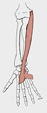
| flexor pollicis longus |
anterior surface of radius and interosseous membrane |
base of the distal phalanx of the thumb |
flexes the metacarpophalangeal and interphalangeal joints of the thumb |
median nerve |
anterior interosseous a. |
the tendon of flexor pollicis longus passes through the carpal tunnel with
the other long digital flexor tendons and the median nerve |
 |
| infraspinatus |
infraspinatous fossa |
greater tubercle of the humerus (middle facet) |
laterally rotates the arm |
suprascapular nerve |
suprascapular a. |
infraspinatus, supraspinatus, teres minor and subscapularis are the rotator
cuff muscles |
 |
| interosseous, dorsal (hand) |
four muscles, each arising from two adjacent metacarpal shafts |
base of the proximal phalanx and the extensor expansion on lateral side
of the 2nd digit, lateral & medial sides of the 3rd digit, and medial side
of the 4th digit |
flex the metacarpophalangeal joint, extend the proximal and distal interphalangeal
joints of digits 2-4, abduct digits 2-4 (abduction of digits in the hand is
defined as movement away from the midline of the 3rd digit) |
ulnar nerve, deep branch |
dorsal and palmar metacarpal aa. |
bipennate muscles; remember DAB & PAD - Dorsal interosseous mm. ABduct and
Palmar interosseous mm. ADduct - then you can figure out where they must insert
to cause these actions |
|
| interosseous, palmar |
four muscles, arising from the palmar surface of the shafts of metacarpals
1, 2, 4, & 5 (the 1st palmar interosseous is often fused with the adductor
pollicis m.) |
base of the proximal phalanx and extensor expansion of the medial side of
digits 1 & 2, and lateral side of digits 4 & 5 |
flexes the metacarpophalangeal, extends proximal and distal interphalangeal
joints and adducts digits 1, 2, 4, & 5 (adduction of the digits of the hand
is in reference to the midline of the 3rd digit) |
ulnar nerve, deep branch |
palmar metacarpal aa. |
unipennate muscles; remember PAD & DAB: Palmar interossei ADduct and Dorsal
interossei ABduct, and you will be able to figure out where they must insert |
|
| latissimus dorsi |
vertebral spines from T7 to the sacrum, posterior third of the iliac crest, lower 3 or 4 ribs, sometimes from the inferior angle of the scapula |
floor of the intertubercular groove |
extends the arm and rotates the arm medially |
thoracodorsal nerve (C7,8) from the posterior cord of the brachial plexus |
thoracodorsal a. |
the inserting tendon twists so that fibers originating highest insert lowest |
 |
| levator scapulae |
transverse processes of C1-C4 vertebrae |
medial border of the scapula from the superior angle to the spine |
elevates the scapula |
dorsal scapular nerve (C5); the upper part of the muscle receives branches of C3 & C4 |
dorsal scapular a. |
levator scapulae is named for its action |
 |
| lumbrical (hand) |
flexor digitorum profundus tendons of digits 2-5 |
extensor expansion on the radial side of the proximal phalanx of digits
2-5 |
flex the metacarpophalangeal joints, extend the proximal and distal interphalangeal
joints of digits 2-5 |
median nerve (radial 2) via palmar digital nerves & ulnar nerve (ulnar 2)
via deep branch |
superficial palmar arterial arch |
lumbricals, (lumbricus is latin for "worm") arise from the profundus tendons
and have the same pattern of innervation as does the profundus muscle (ulnar
and median nn. split the task equally) |
 |
| opponens digiti minimi |
hook of hamate and flexor retinaculum |
shaft of 5th metacarpal |
opposes the 5th digit |
ulnar nerve, deep branch |
ulnar a. |
opposition is a rotational movement of the 5th metacarpal around the long
axis of its shaft; opponens digiti minimi, abductor digiti minimi, and flexor
digiti minimi brevis are in the hypothenar compartment of the hand
|
 |
| opponens pollicis |
flexor retinaculum, trapezium |
shaft of 1st metacarpal |
opposes the thumb |
recurrent branch of median nerve |
superficial palmar branch of the radial a. |
opposition is a rotational movement of the 1st metacarpal around the long
axis of its shaft; opponens pollicis, abductor pollicis brevis, and flexor
pollicis brevis are in the thenar compartment of the hand |
 |
| palmar interosseous |
four muscles, arising from the palmar surface of the shafts of metacarpals
1, 2, 4, & 5 (the 1st palmar interosseous is often fused with the adductor
pollicis m.) |
base of the proximal phalanx and extensor expansion of the medial side of
digits 1 & 2, and lateral side of digits 4 & 5 |
flexes the metacarpophalangeal, extends proximal and distal interphalangeal
joints and adducts digits 1, 2, 4, & 5 (adduction of the digits of the hand
is in reference to the midline of the 3rd digit) |
ulnar nerve, deep branch |
palmar metacarpal aa. |
unipennate muscles; remember PAD & DAB: Palmar interossei ADduct and Dorsal
interossei ABduct, and you will be able to figure out where they must insert |
|
| palmaris brevis |
fascia overlying the hypothenar eminence |
skin of the palm near the ulnar border of the hand |
draws the skin of the ulnar side of the hand toward the center of the palm |
superficial br. of the ulnar n. |
ulnar a. |
palmaris brevis improves the grasp |
 |
| palmaris longus |
common flexor tendon, from the medial epicondyle of the humerus |
palmar aponeurosis |
flexes the wrist |
median nerve |
ulnar a. |
palmaris longus is absent in about 13% of forearms; it may be present on
one side only |
 |
| pectoralis major |
medial 1/2 of the clavicle, manubrium & body of sternum, costal cartilages
of ribs 2-6, sometimes from the rectus sheath of the upper abdominal wall |
crest of the greater tubercle of the humerus |
flexes and adducts the arm, medially rotates the arm |
medial and lateral pectoral nerves (C5-T1) |
pectoral branch of the thoracoacromial trunk |
the deep fascia on its anterior surface should not be fused to the fascia
of the mammary gland - if it is, this is an important clinical sign indicating
breast disease |
 |
| pectoralis minor |
ribs 3-5 |
coracoid process of the scapula |
draws the scapula forward, medialward, and downward |
medial pectoral nerve (C8, T1) |
pectoral branch of the thoracoacromial trunk |
branches of medial pectoral nerve usually pierce pectoralis minor to reach
the pectoralis major muscle |
 |
| pronator quadratus |
medial side of the anterior surface of the distal one-fourth of the ulna |
anterior surface of the distal one-fourth of the radius |
pronates the forearm |
median nerve via the anterior interosseous nerve |
anterior interosseous a. |
pronator quadratus is the deepest muscle in the distal forearm; it works
with pronator teres and has the same nerve supply |
 |
| pronator teres |
common flexor tendon and (deep or ulnar head) from medial side of coronoid
process of the ulna |
midpoint of the lateral side of the shaft of the radius |
pronates the forearm |
median nerve |
ulnar a., anterior ulnar recurrent a. |
median nerve passes between the two heads of origin of pronator teres |
 |
| rhomboideus major |
spines of vertebrae T2-T5 |
medial border of the scapula inferior to the spine of the scapula |
retracts, elevates and rotates the scapula inferiorly |
dorsal scapular nerve (C5) |
dorsal scapular a. |
named for its shape |
 |
| rhomboideus minor |
inferior end of the ligamentum nuchae, spines of vertebrae C7 and T1 |
medial border of the scapula at the root of the spine of the scapula |
retracts, elevates and rotates the scapula inferiorly |
dorsal scapular nerve (C5) |
dorsal scapular a |
named for its shape |
 |
| serratus anterior |
ribs 1-8 or 9 |
medial border of the scapula on its costal (deep) surface |
it draws the scapula forward; the inferior fibers rotate the scapula superiorly |
long thoracic nerve (from ventral rami C5-C7) |
lateral thoracic a. |
a lesion of long thoracic nerve will cause winging of the scapula (i.e.,
the medial border of the scapula falls away from the posterior chest wall and
looks like an angel's wing) |
 |
| serratus posterior inferior |
thoracolumbar fascia, spines of vertebrae T11-T12 and L1-L2 |
ribs 9-12, lateral to the angles |
pulls down lower ribs |
branches of the ventral primary rami of spinal nerves T9-T12 |
lowest posterior intercostal a., subcostal a., first two lumbar aa. |
a respiratory muscle, it receives ventral ramus innervation; embryonically
related to the intercostal muscles, not the deep back mm. |
 |
| serratus posterior superior |
ligamentum nuchae, spines of vertebrae C7 and T1-T3 |
ribs 1-4, lateral to the angles |
elevates the upper ribs |
branches of the ventral primary rami of spinal nerves T1-T4 |
posterior intercostal aa. 1-4 |
a respiratory muscle, it receives ventral ramus innervation; embryonically
related to the intercostal muscles, not the deep back mm. |
 |
| subclavius |
first rib and its cartilage |
inferior surface of the clavicle |
draws the clavicle (and hence the shoulder) down and forward |
nerve to subclavius (C5) |
clavicular br. of the thoracoacromial trunk |
it serves an important protective function - it cushions the subclavian
vessels from bone fragments in clavicular fractures |
 |
| subscapularis |
medial two-thirds of the costal surface of the scapula (subscapular fossa) |
lesser tubercle of the humerus |
medially rotates the arm; assists extention of the arm |
upper and lower subscapular nerves (C5,6) |
subscapular a. |
subscapularis, supraspinatus, infraspinatus, and teres minor are the rotator
cuff muscles |
 |
| supinator |
lateral epicondyle of the humerus, supinator crest & fossa of the ulna,
radial collateral ligament, annular ligament |
lateral side of proximal one-third of the radius |
supinates the forearm |
deep radial nerve |
recurrent interosseous a. |
deep radial nerve passes through the supinator to reach the posterior compartment
of the forearm |
 |
| supraspinatus |
supraspinatous fossa |
greater tubercle of the humerus (highest facet) |
abducts the arm (initiates abduction) |
suprascapular nerve (C5,6) from the superior trunk of the brachial plexus |
suprascapular a. |
supraspinatus initiates abduction of the arm, then the deltoid muscle completes
the action; a member of the rotator cuff group |
 |
| teres major |
dorsal surface of the inferior angle of the scapula |
crest of the lesser tubercle of the humerus |
adducts the arm, medially rotates the arm, assists in arm extension |
lower subscapular nerve (C5,6) from the posterior cord of the brachial plexus |
circumflex scapular a. |
teres major inserts beside the tendon of latissimus dorsi, and assists latissimus
in its actions |
 |
| teres minor |
upper 2/3 of the lateral border of the scapula |
greater tubercle of the humerus (lowest facet) |
laterally rotates the arm |
axillary nerve (C5,6) from the posterior cord of the brachial plexus |
circumflex scapular a. |
fixes the head of the humerus in the glenoid fossa during abduction & flexion
of the arm; a member of the rotator cuff group |
 |
| trapezius |
medial third of the superior nuchal line, external occipital protuberance,
ligamentum nuchae, spinous processes of vertebrae C7-T12 |
lateral third of the clavicle, medial side of the acromion and the upper
crest of the scapular spine, tubercle of the scapular spine |
elevates and depresses the scapula (depending on which part of the muscle
contracts); rotates the scapula superiorly; retracts scapula |
motor: spinal accessory (XI), proprioception: C3-C4 |
transverse cervical a. |
named for its shape; trapezius is an example of a muscle that migrates during
development from its level of origin (cervical) to its final position, pulling
its nerve and artery along behind |
 |
| triceps brachii |
long head: infraglenoid tubercle of the scapula; lateral head: posterolateral
humerus & lateral intermuscular septum; medial head: posteromedial surface
of the inferior 1/2 of the humerus |
olecranon process of the ulna |
extends the forearm; the long head extends and adducts arm |
radial nerve |
deep brachial (profunda brachii) a. |
long head of the triceps separates the triangular and quadrangular spaces
(teres major, teres minor and the humerus are the other boundaries); all three
heads of origin insert by a common tendon |
 |

)
)
)
)
)
)
)
)
)
)
)
)
)
)
)
)
)
)
)
)
)
)
)
)
)
)
)
)
)
)
)
)
)
)
)
)
)
)
)
)
)
)
)
)
)
)
)
)
)
)
)