|
|
Clinical Cases |
|
Abdominal Hernia
Jim is a 53-year-old male, experiencing extreme pain and swelling in the RLQ following the lifting of a heavy pipe. He felt "something tearing" and the pain has not subsided for approximately 40 minuets. Jim has no history of chronic swelling, but did have an appendectomy about 10 years ago. The hernia can be seen as a hypodense area protruding through the anterior abdominal wall from time 0:17-0:18 at J13.
- The most common anterior abdominal wall hernia is: (Please click on the best answer choice below)
- Epigastric (Hernia of Linea Alba)
- Umbilical
- Spigelian
- Incisional
- Subsequent imaging (ultrasound) prompted an immediate trip to the OR for Jim. What was the likely diagnosis? (Please click on the best answer choice below)
- Incarcerated hernia
- Obstructed hernia
- Strangulated hernia
- Most abdominal wall hernias present with moderate pain.
- Umbilical hernias are typically detected during the newborn abdominal examination. They can help to present themselves when the baby cries due to increased intrabdominal pressure. Most, spontaneously resolve due to the natural closure of the umbilical ring. However, if the hernia is large enough it can resist the closure. If this problem persists for 2 years of life or greater, it will likely require surgical intervention. With respect to umbilical hernias, the hernial sac is covered only by skin and subcutaneous tissue? (Please click on the best answer choice below)
- True
- False
A hernia is the protrusion of an organ or the fascia of an organ through the wall of the cavity that normally contains it. An abdominal hernia is the herniation of omentum, intestine, or some other internal body structure through the abdominal wall.
A hernia can be reducible or irreducible. A reducible hernia is one which can be pushed back into place in the abdomen by applying manual pressure to it. Where as, an irreducible hernia cannot be pushed back into place by applying manual pressure. If an abdominal hernia is irreducible there are several potential complications to be aware of. An incarcerated hernia-is one in which adhesions develop between the wall of hernial sac and the wall of the intestine. An obstructed hernia-is one in which the lumen of the herniated part of intestine is obstructed but the blood supply to the hernial sac is intact. A strangulated hernia is one in which the blood supply of/to the sac is cut off, thus, leading to ischemia. The lumen of the intestine may be patent or not. There are several types of internal abdominal and diaphragmatic hernias, however, the focus of this clinical case is on ventral external abdominal wall hernias. These types of hernias can be attributed to either a weak external abdominal wall (via congenital or acquired), or increased intra abdominal pressure (via chronic or acute mechanisms). Ventral hernias occur anteriorly in the abdominal wall and include epigastric (hernia of linea alba), umbilical, Spigelian, parastomal, and most incisional hernias.
Epigastric hernias are defects in the abdominal midline between the umbilicus and the xiphoid process. The defects are often no more than 1 cm in diameter, but can be large and extend from the xiphoid process to the umbilicus. Epigastric hernias can be asymptomatic and can be revealed with increased intra-abdominal pressure (e.g. when patient raises head). The frequency of epigastric hernia is estimated to range from 3 to 5 percent in the general population and is more common in males (male: female = 3:1). It is most commonly diagnosed in middle age. Umbilical hernias occur when part of the intestine protrudes through the umbilical opening in the abdominal muscles. This is a common and typically harmless hernia. They occur most often in infants but can occur in adults as well. In infants if the hernia is large enough it will not allow closure of the umbilical ring, this will require surgical intervention.
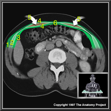
Trunk, Transverse MRI showing Layers of Anterior Abdominal Wall Muscles
- External oblique
- Internal oblique
- Transversus abdominis
- Rectus sheath
- Rectus abdominis
- Linea alba
Image Copyright 1997 The Anatomy Project, Published by Parthenon Publishing Group, Unauthorized use prohibited.
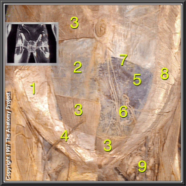
Anterior Abdominal Wall, Lower Part, Dissection
- Internal oblique aponeurosis
- Transversalis fascia
- Rectus abdominis
- External oblique aponeurosis
- Peritoneum
- Inferior epigastric artery and vein
- Arcuate line
- Internal oblique
- Superficial inguinal lymph nodes
Image Copyright 1997 The Anatomy Project, Published by Parthenon Publishing Group, Unauthorized use prohibited.
Lateral ventral hernias are defects at the Spigelian (fascial) zone at any point along its length. This zone is bounded laterally by the muscular fibers of the internal oblique and medially by the lateral margin of the anterior lamina of the rectus sheath.
Incisional hernias, by definition, develop at sites where an incision has been made for some prior abdominal procedure. It is estimated that an incisional hernia will develop in approximately 10 to 15 percent of abdominal incisions, and in up to 23 percent of patients who develop postoperative wound infection.