|
|
Clinical Cases |
|
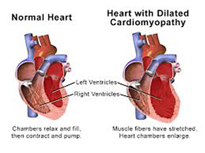
Cardiomegaly is a general term used to describe any condition that results in an enlarged heart. There are two types of cardiomegaly:
- Dilative - The heart can become enlarged due to dilation of the myocardium. An example is Dilated Cardiomyopathy (DCM), which is the most common form of non-ischemic cardiomyopathy. In DCM, the heart becomes weakened and enlarged, and congestive heart failure (CHF) quickly follows. Signs and symptoms are those of left and/ or right heart failure, and signs on autopsy would include central hemorrhagic necrosis in the liver.
Image obtained from the Creative Commons database.
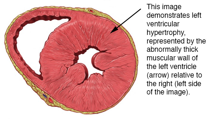
- Hypertrophic - Just as our skeletal muscles hypertrophy (grow in size) in response to increased demand, cardiac muscle undergoes hypertrophy when placed under a high workload for a prolonged period of time. Some cardiac hypertrophy is normal and reversible, such as that seen in athletes and pregnant women. Pathologic hypertrophy is the result of diseases that place increased demand on the heart, such as chronic hypertension, myocardial infarction, and valvular damage. Left ventricular hypertrophy (LVH) is the most common type of hypertrophic heart disease. A common cause of LVH is chronic hypertension, which increases the afterload on the left ventricle. This means the left ventricle has to increase contractility and/ or preload to maintain the same stroke volume. Over time the added stress on the left ventricular myocardium results in muscle hypertrophy and remodeling of the left ventricle to a less efficient size and shape. This leads to a diminishing ejection fraction, meaning the heart must work even harder to maintain cardiac output. The larger heart also demands more blood flow, and so becomes more susceptible to ischemic injury.
Image obtained from the Creative Commons database.
Diagnosis
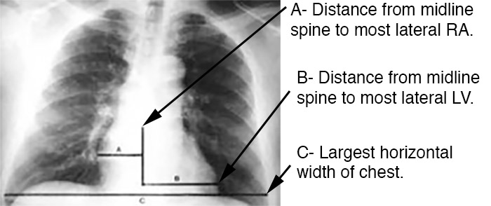
Cardiomegaly is often detected on an anterior-posterior chest x-ray (AP CXR). The standard method for measuring heart size on AP CXR is known as the Danzer Method, and it involves measuring the distance from the midline of the spine to the most lateral aspect of the cardiac apex (distance B, in the image below), and adding this distance to that found from the same midline to the most lateral aspect of the right atrium (distance A). This number is then divided by the largest horizontal width of the chest (distance C), from right to left pleural surface (usually found just above the left hemidiaphragmatic surface). This value (A+B/C) is known as the cardiothoracic ratio (CTR). A CTR > 0.5 indicates cardiomegaly.
Image obtained from the Creative Commons database.
Image obtained from the Creative Commons database.
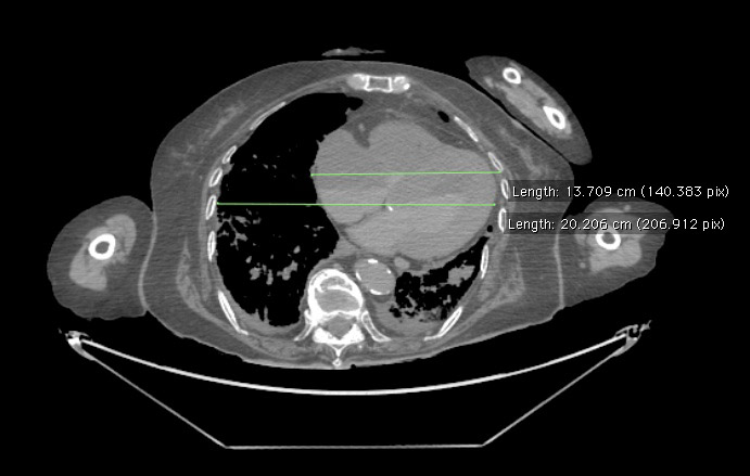
Cardiomegaly can also be diagnosed on CT scan. The screenshot below was taken from the postmortem CT scan of cadaver 33522. Using the medical imaging program Osirix, we measured this patient's CTR and found a heart width of 13.7 cm and a chest width of 20.2 cm, giving us a CTR of 13.7/20.2= 0.68 (this patient's largest horizontal chest width was actually below this cross section, but the difference in width was minimal and so we put both measurements on the same cross-section). Because a CTR less than 0.5 is considered normal, our CT scan indicates that this patient's heart is enlarged.
Image obtained from the Creative Commons database.
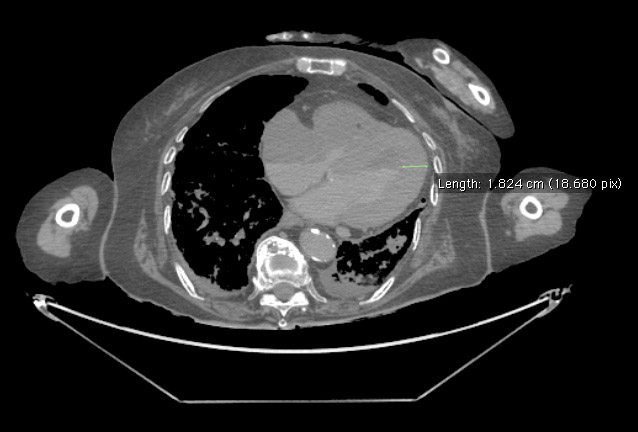
Another measurement was taken of the left ventricular wall thickness, which is normally between 0.6-1.1 cm. As you can in the screenshot below, we measured cadaver 33522's left ventricuar wall thickness to be 1.82 cm, indicating that left ventricular hypertrophy is a component of this patient's cardiomegaly.
Image obtained from the Creative Commons database.