|
|
Clinical Cases |
|
Laryngeal Ossification
A 49-year-old male presents to the clinic with progressive voice changes and dysphagia. A laryngoscopy was performed revealing a small posterior mass that progresses anteriorly. A CT scan shows symmetrical opacities along the thyroid and cricoid. The ossifications (or calcifications) of the larynx are hyperdense lesions seen from time 0:02-0:03 from K4-L4. What is the most likely diagnosis?
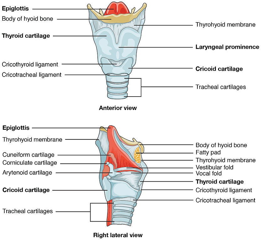
There are three unpaired (thyroid, epiglottis and cricoid) and three paired (arytenoid, cuneiform and corniculate) laryngeal cartilages. The thyroid, cricoid, and greater part of the arytenoid cartilages consist of hyaline cartilage that undergoes calcification and ossification as part of the aging process.
Although the terms calcification and ossification are often used interchangeably they are not the same thing. Calcification precedes ossification when cartilage is transformed into bones. The transition begins posteriorly and progresses anteriorly and superiorly. Ossification is typically first observed in the thyroid, then in the cricoid, and later in the arytenoids. In both males and females, the ossification began at the age of 18 to 20 years in the posterior part of the thyroid cartilage. Ossification of the laryngeal cartilages is usually symmetrical.
This process generally increases with age beginning in the third decade. Typically, the laryngeal cartilages of men will become ossified to a greater extent than those of women. Knowledge of the anatomical location/pattern of cartilage calcification is useful. It can help to differentiate a foreign body from calcification in the thyroid gland. Other anatomical structures to be aware of so as not to confuse with laryngeal ossification are the: styloid process, the stylohyoid ligament, the hyoid bone, vertebral osteophytes, anterior longitudinal ligament, and the tracheal cartilages.