![]()

|
|
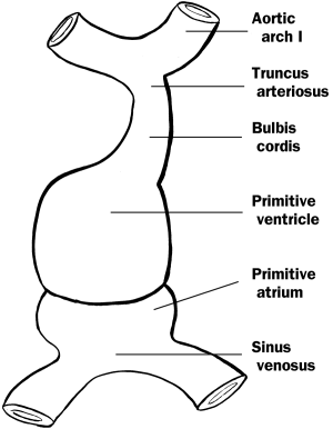
Development of HeartTwo endocardial heart tubes arise from cardiogenic mesoderm. As lateral folding occurs, these fuse to form the primitive heart tube, which develops into the endocardium. The myocardium and epicardium develop from mesoderm surrounding the primitive heart tube. Several contractions and dilations soon appear in the heart tube, all of which have adult remnants. Table 13 - Fates of Embryonic Dilatations of the Primitive Heart Tube
Figure 11 - Primitive heart Development of Blood VesselsBlood vessel formation (angiogenesis) starts at the beginning of the third week. Blood vessels first start to develop in the extraembryonic mesoderm of the yolk sac, connecting stalk, and chorion. Blood vessels begin to develop in the embryo about two days later. Production of BloodProduction of blood (hemopoiesis or hematopoiesis) begins first in the yolk sac wall about the third week of development. Erythrocytes produced in the yolk sac have nuclei. Blood formation does not begin inside the embryo until about the fifth week. Erythrocytes produced in the embryo do not have nuclei (eunucleated). Hematopoiesis inside in the embryo occurs first in the liver, then later in the spleen, thymus, and bone marrow.
Figure 12 - The three embryonic circulations |
|
|