|
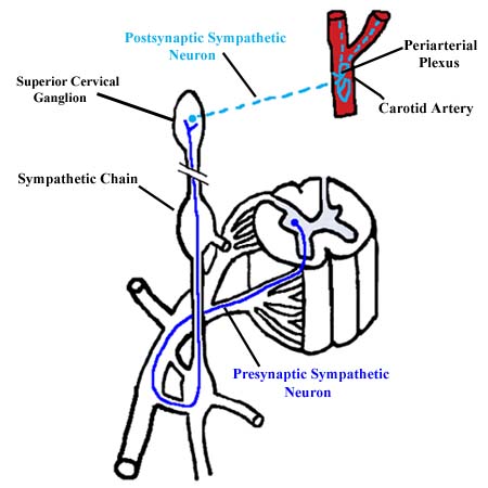
Postsynaptic sympathetic fibers to the neck travel via gray rami to reach ventral primary rami. However, the sympathetic chain ends below the base of the skull as the superior cervical ganglion. So, the cell bodies of the postsynaptic sympathetic fibers to the head are all located in the two (right and left) superior cervical ganglia. Presynaptic sympathetic fibers must ascend through the sympathetic trunk to reach the superior cervical ganglion where they synapse. The postsynaptic sympathetic fibers leave the sympathetic trunk via external and internal carotid nerves that travel to the carotid arteries. These postsynaptic fibers form perivascular plexuses and are distributed to the structures of the head along with the branches of the external and internal carotid arteries. The blood vessels, sweat glands and arrector pili muscles of the head, as well as certain muscles associated with the eye, receive sympathetic innervation. Sympathetic innervation in the head, as with the rest of the body, leads to constriction of the peripheral blood vessels, increase in sweat gland secretion, and contraction of the arrector pili muscles. Sympathetic innervation to the eye results in dilation of the pupil and elevation of the upper eyelid.
|
 |

