Learning Modules - Medical Gross Anatomy
Introduction to Autonomics, Part 2 - Page 8 of 12


Another important difference between the sympathetic and parasympathetic nervous systems is that the presynaptic parasympathetic fibers synapse with terminal ganglion cells that are located individually in or near the wall of the target organ. (This is in contrast to the sympathetic nervous system, in which presynaptic sympathetic fibers synapse in ganglia relatively further away from the target organ.) The exception to this pattern of parasympathetic innervation are 4 pairs of parasympathetic ganglia that occur in the head, although even in the head, these parasympathetic ganglia are relatively close to their targets. So, except for these four pairs of macroscopic ganglia in the head, presynaptic parasympathetic axons travel directly to terminal ganglia (micro-ganglia) within the walls of the target organ.

The presynaptic fibers of the parasympathetic nervous system reach the locations where they synapse by one of three pathways.
1. Most commonly, presynaptic parasympathetic axons travel with their nerves of origin (or in branches that travel into the thorax) directly to ganglia.
2. In the head, some presynaptic parasympathetic axons join the course of an unrelated nerve to arrive at their postsynaptic parasympathetic neuron cell body. In this case, the parasympathetic fibers and unrelated nerve look like one structure and cannot be distinguished.
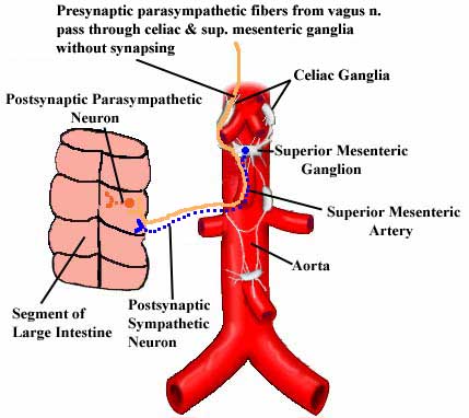
3. Finally, in the abdomen, presynaptic parasympathetic neurons pass through a sympathetic ganglion without synapsing and join a perivascular plexus to form a combined autonomic nerve plexus. They then synapse on cells within the organ wall, like good parasympathetic fibers should.


Go to Question

Quit