|
|
|
||||||||||||
Dissector Answers - Posterior Mediastinum |
|||||||||||||
Learning Objectives and Explanations:
1. Define the boundaries of the posterior mediastinum. (W&B 370-371, N231, TG4-14)2. Describe the major contents of the posterior mediastinum and their relationships. (W&B 370, 408-416, N172, N192, N193, N194, N206, N207, N212, N232, N237, N238, N240, N260, TG4-35, TG4-35, TG4-36, TG4-36, TG4-37, TG4-37)
- Superior: plane b/w sternal angle and T4/T5
- Inferior: diaphragm
- Anterior: pericardium (middle mediastinum)
- Posterior: spinal column
- Vagus nerves (CN X):
- See Objective #1 above.
- Descending aorta (thoracic portion): the continuation of the arch of the aorta, supplying oxygenated blood to thorax (except the heart), abdomen, pelvic region, and lower extremities. It begins on the left side, but moves to the midline (to lie on vertebrae) as it descends. The branches of the thoracic descending aorta include:
- bronchial arteries - supplying the lower trachea and bronchial tree
- pericardial arteries - supplying the pericardium
- posterior intercostal arteries - supplying the intercostal muscles, spinal cord and vertebral column, deep back muscles, and the skin and superficial fascia overlying the intercostal spaces
- superior phrenic arteries - supplying the diaphragm
- esophageal arteries - supplying the lower 2/3 of the esophagus
- mediastinal arteries - supplying the lymph nodes and tissues of the posterior mediastinum
- subcostal arteries - supplying the vertebrae, spinal cord and muscles, skin, and fascia of the upper abdominal wall. They are just like intercostal arteries, but occur below the 12th rib.
Once the thoracic aorta goes through the aortic hiatus in the diaphragm, its name changes to abdominal aorta.
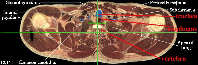
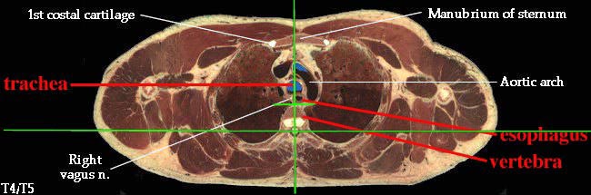
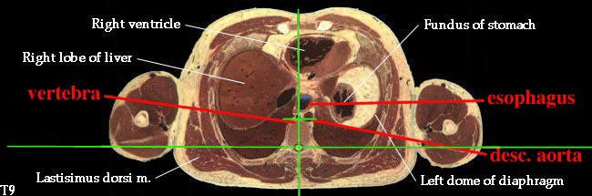
- Esophagus: enters the mediastinum a little to the right of the median plane, posterior to the trachea. It passes to the middle of the body, then to the left and anteriorly as it travels down to the stomach. (See "Extra Explanation" below.)
- Azygos vein system: serves to drain the back, the walls of the thorax and abdomen, and the mediastinal viscera. Although there is a great deal of variation, the usual arrangement is thus (W&B 411-412):
- azygos vein: runs up the right side of the vertebrae, arching over the root of the right lung to join the superior vena cava. The azygos receives blood from the right posterior intercostal veins, as well as the hemiazygos and accessory hemiazygos veins.
- hemiazygos vein: the "inferior" (spatially) partner of the azygos vein. It ascends as far as T9 or so, receiving blood from the left posterior intercostal veins and many of the smaller veins draining the mediastinal viscera, before crossing the vertebral column to join the azygos vein.
- accessory hemiazygos vein: the azygos vein's "superior" partner, running along the left side of the spinal column between T5 and T8. It receives those posterior intercostal veins and others, before crossing the vertebral column to join the azygos vein.
- Thoracic duct: drains all of the lymph from all of the body below the diaphragm and the left half of the body above the diaphragm. In the superior mediastinum it can be found behind the aortic arch, on the left side of the esophagus. (W&B 412)
3. Describe the organization of the thoracic sympathetic trunk, in addition to its visceral and splanchnic branches. (W&B 413-416, N158, N160, N165, N209, N240, N254, TG4-45, TG4-46, TG8-03, TG8-14)This set of cross-sections might help (no guarantee!):
The thoracic sympathetic trunk is the inferior continuation of the cervical sympathetic trunk, and after piercing the diaphragm, continues as the lumbar sympathetic trunk. The trunk is really just a highway for preganglionic sympathetic nerves (thoracolumbar) that need to reach levels superior or inferior to their level of origin in the spinal cord. The chain ganglia along the trunk are the sites of synapse between preganglionic neurons and postganglionic fibers (see box below).
A preganglionic (presynaptic) sympathetic neuron originates in the lateral horn of the spinal cord gray matter. It leaves the cord via the spinal nerve and enters the trunk via a white ramus communicans. The neuron can: a) immediately synapse in the ganglion, or b) travel up or down the trunk, subsequently synapsing in a ganglion superior or inferior to the level of origination of the neuron. In either case, the postganglionic (postsynaptic) neuron then leaves the ganglion via the gray ramus communicans to rejoin the spinal nerve for the purpose of distribution to its target, either via the ventral primary ramus or the dorsal primary ramus.
Besides the three paths mentioned above (up or down via the trunk, or out via the corresponding spinal nerve) there are two other paths that a sympathetic fiber can take. First, some enter visceral branches that directly innervate the smooth muscle, cardiac muscle, and glands of the thoracic viscera. There are contributions to the cardiac, pulmonary, esophageal, and aortic plexuses. Also, there are thoracic splanchnic nerves (Greek, splanchna = viscera), which are preganglionic fibers that travel into and synapse within the abdomen to provide sympathetic innervation for most of the abdominal viscera. (This can be a confusing point. The thoracic splanchnic nerves innervate abdominal viscera. Also, the label "splanchnic" refers to the innveration of viscera, not to a specific origin or make-up. You will see lumbar, pelvic, and sacral splanchnics that are not necessarily pre-ganglionic sympathetic nerves from the sympathetic chain.) These are listed here:
Nerve Level of Origin Site of Synapse great thoracic splanchnic n. T5 - T9 celiac ganglion, superior mesenteric ganglion, suprarenal medulla lesser thoracic splanchnic n. T10 - T11 aorticorenal ganglion least thoracic splanchnic n. T12 renal plexus
Autonomic Nervous System
The autonomic nervous system, made of the parasympathetic nervous system and the sympathetic nervous system, controls all of the smooth muscle in our body, as well as performing some other specialized functions that will be presented in physiology. Smooth muscle is present throughout the body, in blood vessels and skin, but is most often considered in the context of thoracic, abdominal, and pelvic viscera.
In that it innervates muscle, causing contraction or relaxation, the ANS is similar to the system of skeletal muscle innervation. However, the structures of the two systems is different, hence the necessity to learn words like "synapse", and "ganglion". The nerves to skeletal muscle leave the CNS, travel to the muscle, and do their business at the neuromuscular junction. Sympathetic and parasympathetic nerves, however, take a detour along the way. In the ANS, there are two separate neurons that separate the spinal cord from the target, the preganglionic (presynaptic) and the postganglionic (postsynaptic) neurons. What do you suppose is in the middle of the two? Yep, a ganglion. What happens in a ganglion? A synapse.
Preganglionic sympathetic neurons will leave the spinal cord (in the thoracic and lumbar regions) and travel part-way to their target, either via the sympathetic trunk or a splanchnic nerve. Within that trunk or at the end of that splanchnic nerve, there is a ganglion within which the preganglionic sympathetic neuron will synapse with the postganglionic sympathetic neuron. The postganglionic sympathetic neuron will then travel to the target, usually smooth muscle of an arteriole, but also cardiac muscle, sweat glands, and errector pili muscles (small muscles in the skin that attach to hair follicles and can give you "goose bumps").
Preganglionic parasympathetic neurons usually leave the spinal cord (in the cranial and sacral regions) and travel directly to their target organ, sometimes piggybacking on splanchnic or other nerves. They will synapse in very small ganglia in or on the target organ. The postganglionic parasympathetic neurons, then, do not travel very far to reach their target tissue (usually smooth muscle or glands, but also cardiac muscle).
One more thing: You will come across abbreviations, either in the anatomy tables or some texts, like GVE, SVA, GSE, etc. This is old terminology that divided innervation up into general vs. special, visceral vs. somatic, and efferent vs. afferent. The current way of thinking, and the way that this course will be taught, divides innervation up functionally, as described above, with skeletal, sympathetic, and parasympathetic nerves, some of which are efferent and some of which are afferent.
Cultural enrichment: Check out these sections from the 1918 version of Gray's Anatomy of the Human Body! Some of the terms are (of course) out-of-date, but the illustrations are timeless.The Trachea and Bronchi - The Lungs - The Pulmonary Veins - The Thoracic Duct - The Vagus Nerve - Surface Anatomy of the Thorax - Surface Markings of the Thorax
Questions and Answers:
4. Are there sympathetic branches to the lung? Along what do they distribute? (N209, N240, TG4-45, TG4-46, TG8-14)5. Where does the esophagus begin? Where does it pass into the abdomen? Where does it terminate? (N63, N194, N195, N232, N234, N235, TG4-45, TG4-46)Sympathetic fibers reach the lungs via the pulmonary plexuses, which are located along the roots of the lungs. Pulmonary plexuses are continuous with the cardiac plexus at the tracheal bifurcation. Additional sympathetics reach the pulmonary plexuses via the thoracic visceral nerves, which are branches from T1-T4/T5 sympathetic chain ganglia.
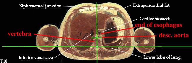
6. Consider the course, relations, constrictions of the esophagus. (N232, TG4-49, TG7-91)The laryngopharynx "becomes" the esophagus at the C6 level. The esophagus passes through the esophageal hiatus of the diaphragm, which is at the T10 level, to end in the cardiac portion of the stomach.
For course and relations, see above. Constrictions of the esophagus are found at its beginning, at the tracheal bifurcation, and at the esophageal hiatus.7. Describe the blood supply and venous drainage of the esophagus. Are there venous collaterals to stomach? (N238, TG4-49, TG7-91)8. Do the right and left mediastinal pleurae come together? (N194, TG4-49)Two or three esophageal arteries branch from the descending aorta. Esophageal veins drain into the azygos system, which eventually reaches the superior vena cava. The esophageal tributaries of the left gastric vein drain the terminal esophagus. Since the gastric veins first drain into the portal system before going to the heart, this part of the esophagus is an important site of portal-caval (portal-systemic) anastomosis in cases of portal hypertension.
9. Through what and at what level does the aorta enter the abdominal cavity? (N194, N195, TG4-37, TG4-38, TG4-39)Very low in the posterior mediastinum the esophagus sweeps forward, so there is potential for the right and left mediastinal parietal pleurae to touch one another posterior to esophagus and anterior to aorta. However, typically the anterior deviation of the esophagus is not sufficient to allow enough space for this contact of the pleurae.
10. What is the subcostal artery? (N264, TG4-49, TG7-91, TG4-39)The descending thoracic aorta passes through the diaphragm at the aortic hiatus, a passageway between the two diaphragmatic crura, located at the T12 level.
11. Completely review the blood supply to an intercostal space. (N33, N191, N192, TG4-43)Below the 12th rib, there is no intercostal space or intercostal artery, so we call the segmental neurovascular structures subcostal.
12. What are the posterior branches of the posterior intercostal (segmental) arteries? What do they supply? (N171, N172, TG4-39, TG4-08)Intercostal spaces in general are supplied by posterior and anterior intercostal arteries. Posterior intercostal arteries 3 through 11 are branches of the descending thoracic aorta. The first two posterior intercostal arteries are branches of the highest intercostal artery, which is a branch of the costocervical trunk from subclavian artery. Anterior intercostals are branches of the internal thoracic or the musculophrenic arteries.
13. Observe the azygos venous system. If you have two primary veins, do they communicate with one another? How? Where? What is the pattern of venous drainage in your specimen? Are all the veins present? If not, where does the drainage go? (N238, TG4-38, TG4-39, TG4-40)The posterior branches of posterior intercostal arteries supply the deep and superficial back muscles, skin of the back, and the vertebral column. They have radicular branches that reach the spinal cord along the dorsal and ventral rootlets.
14. What vein drains the first intercostal space? Into what? What veins drain into the azygos system? (N238, TG4-38, TG4-39, TG4-40)Hemiazygos and accessory hemiazygos usually cross the midline at T7, 8, or 9 to empty into the azygos vein. There are also connections between the various left-sided venous channels.
15. How does the thoracic duct get into the thorax? At what level does it deviate to the left side? (N206, N266, TG4-43, TG4-44)Both first posterior intercostal veins drain directly into their respective brachiocephalic veins. Bronchial and esophageal veins drain into the azygos system, the latter being a significant site of portal-caval anastomosis in cases of portal hypertension. (This is the second mention... could be important!)
16. Do you find posterior mediastinal lymph nodes or afferent lymph channels? (N239, TG4-43, TG4-44)The thoracic duct enters the chest through the aortic hiatus along the right side of the aorta. It deviates to the left at the level of the sternal angle. (The trachea pushes the esophagus against the vertebral bodies which pushes thoracic duct to the left.)
17. What are bronchomediastinal lymph trunks? (N208, TG4-43, TG4-44)The thoracic duct is usually paralleled by posterior mediastinal nodes.
18. Is the sympathetic trunk located within the posterior mediastinum? Does it change positions in different regions of the chest? (N N180, N254, TG4-45, TG4-46, TG8-14)The lymph from the lungs and chest passes through the paratracheal nodes to form the bronchomediastinal lymph trunks. These drain to the thoracic duct on the left and the right lymphatic duct on the right.
19. How many thoracic ganglia do you find? (N209, N240, TG4-45, TG4-46, TG8-14)The sympathetic trunk lies on the heads of the ribs through most of the chest, so it is almost, but not quite, within the posterior mediastinum. It deviates anteromedially as it travels inferiorly to its exit through the diaphragm.
20. Identify white and gray rami communicans. What is their significance and distribution? What do they contain? (N164, N180, N209, N240, N254, TG4-45, TG4-46, TG8-14)There are typically 12 thoracic ganglia, although the first may be fused with the inferior cervical ganglion to form a cervicothoracic (stellate) ganglion.
21. Do you see thoracic visceral nerves to the aorta, esophagus, and trachea? What about to the cardiac and pulmonary plexuses? (N209, N240, TG4-45, TG4-46, TG8-03, TG8-14)Since the preganglionic sympathetic neurons live within the spinal cord at T1 to L2 levels, these are the only levels where white rami communicans are found. Gray rami, on the other hand, are found at every level at which there are spinal nerves. Gray rami carry postganglionic sympathetic fibers back to the ventral primary rami, to be distributed along their branches (and also the branches of the dorsal primary rami).
22. Expose the greater (thoracic) splanchnic nerve. From what does it come? At what level? What types of fibers does it contain? To what does it distribute? (N209, TG4-45, TG4-46, TG8-03, TG8-14)The first 4 or 5 thoracic ganglia (T1-4) give small visceral branches that pass anteroinferiorly to reach the cardiac, pulmonary, esophageal, and aortic plexuses, as well as the trachea.
The greater thoracic splanchnic nerve is made by contributions from sympathetic chain ganglia at T5 to T9 (or T10) levels. These are preganglionic fibers that leave the chest to enter the abdomen. They synapse in the celiac ganglion and innervate the abdominal viscera that is supplied by the celiac trunk.