|
|
|
||||||||||||
Lab Manual - Forearm & Wrist |
|||||||||||||
Assignments:
- Before lab:
- Download and review the Lab Overview PowerPoint.
- Review the Prelab Learning Module.
- Review the Steps of Dissection and Dissection Videos.
- During lab:
- Follow the steps of the dissection procedure in the Lab Manual (this page).
- Be certain to identify all of the Review Items.
- After lab:
- Read the Dissector Answers to cover the Learning Objectives for this lab.
- Read the Clinical Case for this lab.
- Review the Practice Questions for this lab.
Learning Objectives:
Upon completion of this session, the student will be able to:
- Identify the prominent features of the humerus, ulna, radius, carpals, metacarpals and phalanges of the associated extensor and flexor compartments as given in the lab manual.
- Identify the extensor and flexor compartments of the forearm and hand, the nerve and vessels supplying their contents, and the functional significance of the included muscles.
- Correlate any fractures or deep cuts of the forearm or hand with functional disruptions of associated muscular or neurovascular structures.
- Describe the movements of elbow, wrist, and finger joints.
- Identify position of tendons and associated bursae beneath the extensor retinaculum and palmar carpal ligament.
Procedure:
1. Review the bony landmarks. (Play movie; View images: N 420, 421, 436, 439, 440, 450, 451, 452, 456, TG 2-03A, 2-03B, 2-04, 2-21, 2-22, 2-31, 2-32)
Review bony processes of the humerus, radius and ulna previously examined. Identify supinator crest, posterior border, and interosseous crest of ulna. Identify the dorsal radial tubercle and interosseous crest of radius. Locate grooves at the distal end of radius which accommodate tendons as they cross the wrist. Examine an articulated hand, noting arrangements of carpals, metacarpals, and phalanges.
Identify proximal, middle and distal phalanges.
Bones of the elbow Bones of the elbow Bones of the forearm Bones of the forearm Bones of the wrist Wrist bones 2. Remove the superficial fascia from the dorsum of the forearm. (Play movie; View images: N 444, 480, TG 2-02, 2-23, 2-29)
Remove the superficial fascia from the forearm, wrist and hand, leaving the cutaneous nerves and veins intact. Observe the posterior part of the antebrachial fascia, noting continuity with brachial fascia and its thickening at the distal end of radius and ulna and wrist, the extensor retinaculum. Note its continuation onto the hand as the fascia of the dorsum of the hand. Remove the posterior antebrachial fascia leaving the extensor retinaculum intact. As you reflect, note that some muscles use this fascia as an origin. Do you see intermuscular septa? If not, why?
3. On the extensor side of the forearm, separate and dissect the superficial extensor muscles. (Play movie; View images: N 444, 446, 450, 451, TG 2-21, 2-22, 2-23, 2-29)
The muscles of the posterior forearm region lie in two layers, superficial and deep. The superficial layer arises from the lateral epicondyle of the humerus via a common extensor tendon. The muscles of the deep layer arise collectively from the posterior surface of the radius and ulna and the intervening interosseous membrane. Emphasize these general common origins. Note functional arrangements of muscles. Note that muscles use adjacent muscles as part of their origin.
Dissect each muscle by picking up its tendon at the proximal border of the extensor retinaculum and separating it toward its origin as far as possible, in the following order: brachioradialis (note attachment to lateral supracondylar ridge), extensor carpi radialis longus (note attachment to lateral supracondylar ridge), extensor carpi radialis brevis, extensor digitorum, extensor digiti minimi, and extensor carpi ulnaris.
4. Examine extensor expansion on dorsum of fingers. (Play movie; View images: N 441, 451, 463, 464, 465, 470, TG 2-22, 2-32, 2-34, 2-35, 2-40, 2-41A, 2-41B, 2-45)
Remove the deep fascia from the dorsum of the hand distal to the extensor retinaculum. Identify tendons of extensor digitorum and extensor digiti minimi muscles. Trace an exemplary tendon to any digit (2-4) and examine the manner of insertion on the middle and distal phalanges. Expose completely an extensor expansion, demonstrating its lateral borders, interossei and lumbrical tendons. Do not trace these tendons into the palm. Note transverse fibers uniting all tendons and capsular attachments at the joints. Note tendinous cross bridges.
Return to the wrist and locate insertions of the extensor carpi radialis longus and brevis and extensor carpi ulnaris.
5. Dissect the deep extensor muscles of the forearm. (Play movie; View images: N 439, 441, 444, 445, 447, 451, 452, 454, 456, 459, 460, 461, 462, 468, 470, 472, 476, 478, TG 2-24, 2-27, 2-29, 2-30, 2-31, 2-32A, 2-32B, 2-34, 2-35, 2-39A, 2-39B, 2-40A, 2-40B, 2-41, 2-50)
Return to the forearm and make an incision between the extensor carpi radialis brevis and extensor digitorum as far proximal as the lateral epicondyle of the humerus. Separate these muscles to expose the deep layer.
Clean and identify the following muscles: supinator, abductor pollicis longus, extensor pollicis brevis, extensor pollicis longus, and extensor indicis. Trace the tendons to the extensor retinaculum and then, distal to the extensor retinaculum, to their ultimate insertions. Note the course of some of these tendons across the tendons of extensor carpi radialis longus and brevis.
Examine the extensor retinaculum, noting medial and lateral attachments, proximal and distal extents, and orientation. Open each compartment with a longitudinal incision and note: (1) tendons contained, (2) deep attachments of septa, (3) relation of tendons to grooves in bone, (4) synovial bursae (sheaths), (5) sequence of tendons (from radial to ulnar side).
Synovial bursa (sheath). Carefully open a compartment and distinguish parietal and visceral layers. Where do they become continuous?
Radial nerve. Return to the cubital fossa. Locate the radial nerve beneath the brachioradialis muscle. Identify branches of the radial nerve to forearm muscles. Note point of division into superficial and deep radial nerves. Follow the superficial radial nerve, noting course and relations to brachioradialis, and point of emergence into the superficial fascia. What does it accompany? Follow the deep radial nerve as it pierces the supinator muscle. Trace by incising through the muscle. Observe branches of the deep radial nerve to supinator and superficial extensor muscles. Trace its branches to the muscles of the deep layer. What is the posterior interosseous nerve?
Find and save the dorsal cutaneous branch of the ulnar nerve as it passes around the distal end of the ulna onto the dorsum of the hand. Identify its dorsal digital branches.
Review radius and ulna and general arrangement of carpals, metacarpals, and phalanges in an articulated hand.
Surface anatomy of the anatomical snuffbox Examine the anterior part of the antebrachial fascia and, in its distal part, the palmar carpal ligament spanning between the distal end of the radius and ulna. Note differences between this and the extensor retinaculum.
6. On flexor side of forearm, separate and dissect the various muscular layers and trace the median and ulnar nerves. (Play movie; View images: N 434, 445, 446, 447, 448, 449, 450, 451, 460, 461, 465, 466, 468, 472, 475, 476, 481, TG 2-19, 2-21, 2-22, 2-23, 2-24, 2-25, 2-27A, 2-27B, 2-28B, 2-28C2-30, 2-33, 2-34, 2-36, 2-37A, 2-37B2-38, 2-39, 2-40, 2-41, 2-48, 2-49A, 2-49B, 2-51A, 2-51B)
The forearm flexor muscles are arranged in three layers. Dissect each layer separately.
LAYER 1. Pronator teres. Note two heads of origin, relation of median nerve. What structures cross its insertion?
Pronation of the hand LAYER 2. Flexor digitorum superficialis. Trace its origin along the oblique line of the radius to the coronoid process of the ulna and to the medial epicondyle of the humerus. Note the fibrous arch spanning between radius and ulna.
LAYER 3. Incise the radial origin of flexor digitorum superficialis and reflect. Dissect flexor pollicis longus, flexor digitorum profundus and pronator quadratus (deep to the tendons of the above muscles). Trace tendons to the wrist but do not invade the palm. Compare the arrangement of the tendons of flexor digitorum superficialis and profundus as they enter the wrist. Note the anterior interosseous artery, vein and nerve.
Return to the cubital fossa and trace the brachial artery as it divides into radial and ulnar arteries. Where is the point of bifurcation?
NOTE specifically the relations at the wrist between the radial artery and the flexor carpi radialis and the brachioradialis in the palmar carpal ligament. This is a pulse point. Palpate on your partners. The grouping of the radial artery and superficial radial nerve under cover of brachioradialis constitutes a convenient dividing line between forearm flexor and extensor muscle groups.
Follow the ulnar artery, noting course and specific relations to median and ulnar nerves and to muscles. Locate it at the wrist between the flexor carpi ulnaris and flexor digitorum superficialis, deep to the palmar carpal ligament. This is a pulse point; palpate on your partners. Identify the following branches: common interosseous, anterior interosseous, posterior interosseous; note the muscular branches. Consider the collateral circulation of the elbow between branches of brachial, deep brachial, radial and ulnar arteries. Note the accompanying veins (venae comitantes). Consider the blood supply to the entire forearm.
Angiogram of collateral vessels Median nerve. Trace through the forearm, noting specific relations to pronator teres, the fibrous arch of flexor digitorum superficialis, the ulnar artery, its location between flexor digitorum superficialis and profundus. Where do muscular branches to the flexor muscles arise?
Ulnar nerve. Trace through the forearm; note specific relations to flexor carpi ulnaris, to flexor digitorum superficialis and profundus, and to the ulnar artery. Precisely locate at the wrist. What muscles does it supply? Where do these branches leave the ulnar nerve?
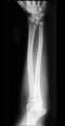
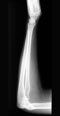
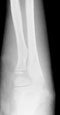
Elbow dislocation Elbow dislocation Growth plates