|
|
|
||||||||||||
Dissector Answers - Forearm & Wrist |
|||||||||||||
Learning Objectives:
Upon completion of this session, the student will be able to:
- Identify the prominent features of the humerus, ulna, radius, carpals, metacarpals and phalanges of the associated extensor and flexor compartments as given in the lab manual.
- Identify the extensor and flexor compartments of the forearm and hand, the nerve and vessels supplying their contents, and the functional significance of the included muscles.
- Correlate any fractures or deep cuts of the forearm or hand with functional disruptions of associated muscular or neurovascular structures.
- Describe the movements of elbow, wrist, and finger joints.
- Identify position of tendons and associated bursae beneath the extensor retinaculum and palmar carpal ligament.
Learning Objectives and Explanations:
1. Identify the prominent features of the humerus, ulna, radius, carpals, metacarpals and phalanges of the associated extensor and flexor compartments as given in the lab manual. (W&B 127-129, 143-145, 154-156, N420, N421, N436, N439, N440, N450, N451, N452, N456, TG2-04, TG2-31, TG2-32)2. Identify the extensor and flexor compartments of the forearm and hand, the nerve and vessels supplying their contents, and the functional significance of the included muscles. (W&B 135-143, N434, N439, N441, N444, N445, N446, N447, N448, N449, N450, N451, N452, N454, N456, N459, N460, N461, N462, N463, N464, N465, N466, N468, N470, N472A, N472B, N475, N476, N478, N481, TG2-23, TG2-24, TG2-25, TG2-26A, TG2-26B, TG2-27A, TG2-27B, TG2-28B, TG2-28C, TG2-29, TG2-30)
- humerus: the bone of the upper arm. Its parts also serve as the origin for many muscles of the forearm. The medial epicondyle is the attachment site for the common flexor tendon, which gives rise to the superficial group of forearm flexor muscles (See #2 below). The lateral epicondyle is the attachment site for the common extensor tendon, which is the origin of some forearm extensor muscles (See #2 below). The lateral supracondylar ridge gives rise to the brachioradialis muscle and the extensor carpi radialis longus muscle.
- ulna: the medial bone of the forearm. It is more firmly connected to the humerus than the radius, but it is only indirectly articulated with the wrist and hand. Note that the head of the ulna is located distally. The tuberosity of the ulna is a point of insertion for the brachialis tendon along with the coronoid process, which also forms part of the trochlear notch.
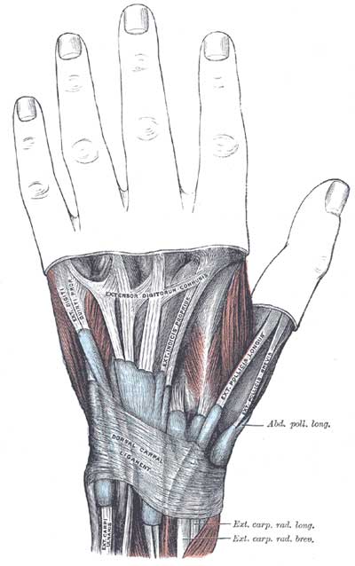
- radius: the shorter, laterally-placed bone of the forearm. Its head is located on its smaller proximal end, and its lower end broadens to take almost the full contact of the bones of the wrist. The tuberosity of the radius is the insertion point for the biceps brachii tendon. The interosseous crest is a point of attachment for the interosseous membrane, and the dorsal radial tubercle acts as a pulley point for the tendon of extensor pollicis longus, separating it from the tendons of the extensor carpi radialis longus and brevis muscles.
- carpals: These eight small bones of the wrist are held together by ligaments and arranged in two rows, proximal and distal. The bones of the proximal row, listed from the radial to the ulnar side, are the scaphoid, the lunate, the triquetrum, and the pisiform. In the distal row, from radial to ulnar side, are the trapezium, the trapezoid, and capitate, and the hamate. Read across the proximal layer of bones: Send Louis To Paris. Read across the distal layer of bones: To Tame Carnal Hunger. (Note that the pisiform bone is a sesamoid bone in the tendon of the flexor carpi ulnaris.)
- metacarpals: There are five metacarpal bones, numbered from 1 (the thumb) to 5 (the little finger). These bones are just distal to the carpals.
- phalanges: There are fourteen of these "bones of the fingers." The thumb has only two phalanges, a proximal and distal, where as the other digits each have three phalanges, proximal, middle, and distal.
3. Correlate any fractures or deep cuts of the forearm or hand with functional disruptions of associated muscular or neurovascular structures. (N461,N466,N472,N475,N477,N478N481, TG2-23, TG2-24, TG2-25, TG2-26A, TG2-26B, TG2-27A, TG2-27B, TG2-28B, TG2-28C, TG2-29, TG2-30)The forearm is organized into anterior and posterior compartments separated by the interosseous membrane that connects the radius and ulna. The anterior compartment contains the flexor muscles, together with the median nerve (and branches), the ulnar nerve, and accompanying vessels. The posterior compartment contains the extensor muscles (with the exception of the brachioradialis, which is an elbow flexor), the radial nerve, and its branches. There are nineteen muscles in the forearm. Within both the posterior and anterior compartments there are two and three layers of muscle groups, respectively. A good way to memorize the muscles is by group level. The tables immediately below are listed according to that. Further below is a list of the muscles sorted by function.
superficial extensors:
Muscle Origin Insertion Action Innervation Blood Supply brachioradialis upper two-thirds of the lateral supracondylar ridge of the humerus lateral side of the base of the styloid process of the radius flexes the elbow, assists in pronation & supination radial nerve radial recurrent a. extensor carpi radialis longus lower one-third of the lateral supracondylar ridge of the humerus dorsum of the second metacarpal bone (base) extends the wrist; abducts the hand radial nerve radial a. extensor carpi radialis brevis common extensor tendon (lateral epicondyle of humerus) dorsum of the third metacarpal bone (base) extends the wrist; abducts the hand deep radial nerve radial a.
extensor digitorum common extensor tendon (lateral epicondyle of the humerus) extensor expansion of digits 2-5 extends the metacarpophalangeal, proximal interphalangeal and distal interphalangeal joints of the 2nd-5th digits; extends wrist deep radial nerve interosseous recurrent a. and posterior interosseous a. extensor digiti minimi common extensor tendon (lateral epicondyle of the humerus) joins the extensor digitorum tendon to the 5th digit and inserts into the extensor expansion extends the metacarpophalangeal, proximal interphalangeal and distal interphalangeal joints of the 5th digit deep radial nerve interosseous recurrent a. extensor carpi ulnaris common extensor tendon & the middle one-half of the posterior border of the ulna medial side of the base of the 5th metacarpal extends the wrist; adducts the hand deep radial nerve ulnar a.
deep extensors:
Muscle Origin Insertion Action Innervation Blood Supply supinator lateral epicondyle of the humerus, supinator crest & fossa of the ulna, radial collateral ligament, annular ligament lateral side of proximal one-third of the radius supinates the forearm deep radial nerve recurrent interosseous a. abductor pollicis longus middle one-third of the posterior surface of the radius, interosseous membrane, mid-portion of posterolateral ulna radial side of the base of the first metacarpal abducts the thumb at carpometacarpal joint deep radial nerve posterior interosseous a. extensor pollicis brevis interosseous membrane and the posterior surface of the distal radius base of the proximal phalanx of the thumb extends the thumb at the metacarpophalangeal joint deep radial nerve posterior interosseous a extensor pollicis longus interosseous membrane and middle part of the posterolateral surface of the ulna base of the distal phalanx of the thumb extends the thumb at the interphalangeal joint deep radial nerve posterior interosseous a extensor indicis interosseous membrane and the posterolateral surface of the distal ulna its tendon joins the tendon of the extensor digitorum to the second digit; both tendons insert into the extensor expansion extends the index finger at the metacarpophalangeal, proximal interphalangeal and distal interphalangeal joints deep radial nerve posterior interosseous a
superficial flexors:
Muscle Origin Insertion Action Innervation Blood Supply pronator teres common flexor tendon and (deep or ulnar head) from medial side of coronoid process of the ulna midpoint of the lateral side of the shaft of the radius pronates the forearm median nerve ulnar a., anterior ulnar recurrent a. flexor carpi radialis common flexor tendon from the medial epicondyle of the humerus base of the second and third metacarpals flexes the wrist, abducts the hand median nerve ulnar a. palmaris longus common flexor tendon from the medial epicondyle of the humerus distal half of flexor retinaculum and palmaris aponeurosis flexes hand (at wrist) and tightens palmar aponeurosis median nerve (C7 and C8) ulnar a. flexor carpi ulnaris common flexor tendon & (ulnar head) from medial border of olecranon & upper 2/3 of the posterior border of the ulna pisiform, hook of hamate, and base of 5th metacarpal flexes wrist, adducts hand ulnar nerve ulnar a. flexor digitorum superficialis humeroulnar head: common flexor tendon; radial head: middle 1/3 of radius shafts of the middle phalanges of digits 2-5 flexes the metacarpophalangeal and proximal interphalangeal joints median nerve ulnar a.
deep flexors:
Muscle Origin Insertion Action Innervation Blood Supply flexor digitorum profundus posterior border of the ulna, proximal two-thirds of medial border of ulna, interosseous membrane base of the distal phalanx of digits 2-5 flexes the metacarpophalangeal, proximal interphalangeal and distal interphalangeal joints median nerve (radial one-half) via anterior interosseous n.; ulnar nerve (ulnar one-half) ulnar a., anterior interosseous a. flexor pollicis longus anterior surface of radius and interosseous membrane base of the distal phalanx of the thumb flexes the metacarpophalangeal and interphalangeal joints of the thumb median via anterior interosseous n. anterior interosseous a.
pronator quadratus medial side of the anterior surface of the distal one-fourth of the ulna anterior surface of the distal one-fourth of the radius pronates the forearm median via anterior interosseous n. anterior interosseous a.
Sorted by function:
flexor division:extensor division:
- muscles which rotate the radius on the ulna:
- pronator teres (superficial group, anterior compartment - pronates)
- pronator quadratus (deep group, anterior compartment - pronates)
- supinator (deep group, posterior compartment - supinates)
- muscles which flex the hand at the wrist:
- flexor carpi radialis (superficial group, anterior compartment)
- flexor carpi ulnaris (superficial group, anterior compartment)
- palmaris longus (superficial group, anterior compartment)
- muscles which flex the digits:
- flexor digitorum superficialis (intermediate group, anterior compartment)
- flexor digitorum profundus (deep group, anterior compartment)
- flexor pollicis longus (deep group, anterior compartment)
- muscles which extend the hand at the wrist:
- extensor carpi radialis longus (superficial group, posterior compartment)
- extensor carpi radialis brevis (superficial group, posterior compartment)
- extensor carpi ulnaris (superficial group, posterior compartment)
- muscles which extend the digits, except the thumb:
- extensor digitorum (superficial group, posterior compartment)
- extensor indicis (deep group, posterior compartment)
- extensor digiti minimi (superficial group, posterior compartment)
- muscles which operate in extension or abduction of the thumb:
- abductor pollicis longus (deep group, posterior compartment - abducts thumb)
- extensor pollicis brevis (deep group, posterior compartment)
- extensor pollicis longus (deep group, posterior compartment)
4. Describe the movements of elbow, wrist, and finger joints. (N436,N439A,N439B,N453A,N453B,N453C,N458, TG2-43A, TG2-43BC, TG2-44A, TG2-44B, TG2-44C, TG2-45A, TG2-45B)
- The flexor digitorum superficialis has four tendons, which at the wrist lie in two layers. The tendons destined for the third and fourth fingers lie superficially in the compartment; the second and fifth digits are more deeply placed. As a result, a deep cut to the wrist would more likely leave the third and fourth flexor tendons injured. (Note that a cut to this part of the wrist, a frequently attempted spot for suicide, would be more likely to cause injury to cutaneous veins or flexor tendons 3 and 4 than to an artery. No major artery is located at this position.)
- The radial nerve may be injured in its groove on the posterior aspect of the humerus. By this level the nerve has given off all its branches to the triceps muscle so there is no loss of extension at the elbow. However, all postaxial muscles (forearm extensors) below this level would be paralyzed, resulting in "wrist-drop" and the inability to extend the hand or digits. (The interphalangeal joints can still be extended by the unaffected interosseus and lumbrical muscles.) There will be sensory loss in the areas served by the posterior antebrachial cutaneous and superficial radial nerves.
- Injury to the median nerve at the wrist emphasizes the importance of opposition in the activities of the hand. Loss of the motor or recurrent branch of the median nerve paralyzes the muscles of the thenar eminence, with subsequent wasting of this area. The thumb can no longer be opposed to the other digits and the normal grasp of the hand is lost. There is also important sensory loss in the areas of distribution of the proper digital branches of the median nerve to the digits.
- Injury to the ulnar nerve at the wrist produces a deformed "claw-hand". The thumb is strongly abducted and all the metacarpophalangeal joints are hyperextended. There is marked wasting through the hand, as well as sensory defects in the little finger and medial half of the ring finger.
5. Identify position of tendons and associated bursae beneath the extensor retinaculum and palmar carpal ligament. (W&B 149-154, N461, N462, N463,N470, TG2-40A, TG2-41A, TG2-34B)
- The wrist joint, also called the radiocarpal articulation, has great movement ability because of its convex oval articular surface. The joint can flex, extend, abduct, adduct and circumduct. Rotary motion is prohibited.
- The "knuckles", or metacarpophalangeal joints (MP), are characterized by loose articular capsules. Movements of flexion and extension, abduction and adduction and circumduction are permitted at these joints. Extension is making a flat hand; flexion is making a fist. The metacarpophalangeal joint of the thumb is limited to the actions of flexion and extension. (The thumb's freedom of movement is a result of its carpometacarpal joint).
- The elbow joint is essentially a hinge joint (ginglymus). However, the elbow joint also includes within a common articular capsule the proximal radioulnar joint, an articulation which is described with other radioulnar articulations. The movements of the elbow joint are flexion and extension. The freedom with which the hand can be elevated in flexion at the elbow is due to the slight medial rotation of the humerus and the semipronated position of the forearm, which is habitual. Flexion of the elbow joint is produced by the action of the biceps and brachialis muscles with the assistance of brachioradialis and those forearm muscles arising from the medial epicondyle (common flexor tendon). Extension of the elbow is due to the pull of the triceps and anconeus muscles. Please refer to Woodburne and Burkel pages 174-5 for good diagrams and a complete description of this joint.
Images from "Anatomy of the Human Body" by Henry Gray are provided by:
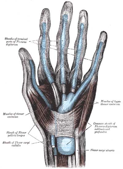
Synovial sheaths, like bursae, reduce the frictional effects of the passage of the tendons through tight compartments of the wrist. A sheath is formed like a double-walled tube, the delicate inner wall closely attached to the tendon and its outer wall lining the compartment in which the tendon lies. The two layers are continuous with one another at the ends of the tube.
Each of the compartments on the dorsum of the wrist contains a synovial sheath investing the tendon(s) included in the compartment. The upper ends of the sheaths on the dorsum of the hand lie at the upper border of the extensor retinaculum, and they extend variable distances distal to it. They end just short of the inserting tendon (For example, the extensor carpi radialis longus and brevis, and the extensor carpi ulnaris sheaths end just short of their insertions on the bases of the metacarpal bones.)
On the palmar aspect of the wrist the tendons of the palmaris longus and flexor carpi ulnaris are not provided with synovial sheaths. The digital flexors, however, are protected at the wrist by complex synovial coverings: The radial bursa is a long synovial sheath for the tendon of flexor pollicis longus, which extends along this tendon from several centimeters above the flexor retinaculum to just proximal to its insertion on the distal phalanx of the thumb. The ulnar bursa is the complex covering of the digital flexor tendons. It occupies the center of the fibro-osseous tunnel of the wrist and also extends above the flexor retinaculum for several centimeters. This sheath exhibits the invaginated character of synovial sheaths. The general sheath for these eight tendons continues to about the middle of the palm and terminates there, except for the portion concerned with the fifth digit, which continues as far as the insertion of the profundus tendon on the base of the distal phalanx.
Cultural enrichment: Check out these sections from the 1918 version of Gray's Anatomy of the Human Body! Some of the terms are (of course) out-of-date, but the illustrations are timeless. The Anterior Divisions (nerves) - The Veins of the Upper Extremity and Thorax - The Brachial Artery - The Radial Artery - The Ulnar Artery - The Muscles and Fascia¾ of the Forearm - Surface Anatomy of the Upper Extremity - Surface Markings of the Upper Extremity
Questions and Answers:
6. After removing the posterior antebrachial fascia (leaving the extensor retinaculum intact), do you see intermuscular septa? If not, why?The answer is no. At this distal part of the forearm, intermuscular septum would limit movement of the muscles contained.6a. Carefully open a compartment and distinguish parietal and visceral layers. Where do they become continuous?The parietal layer of a synovial sheath reflects onto the visceral layer (on the associated tendon) both proximally and distally to its passage, deep to a retinaculum. (N462, TG2-40A, TG2-41A)6b. How do bursae function?Bursae and synovial sheaths are synovial bags containing synovial fluid, which gives these bags a lubricating quality, reducing the friction on tendons.6c. What is the posterior interosseous nerve?The posterior interosseous nerve is the sensory continuation of the deep radial nerve, distal to its motor branches to the extensor muscles. It reaches the wrist joint and carpal bones for proprioceptive sense from these structures. (N445,N478, TG2-30)7. Review the radius, ulna, and the general arrangement of carpals, metacarpals, and phalanges in an articulated hand.See #1 above. (N436,N439,N452,N456, TG2-04, TG2-31, TG2-32)8. Note the differences between the palmar carpal ligament and the extensor retinaculum.The extensor retinaculum extends from the lateral margin of the radius to the styloid process of the ulna, the pisiform bone, and the triquetrum. The retinaculum has deep attachments to the ridges on the dorsum of the distal end of the radius. The palmar carpal ligament is also a thickening of the antebrachial fascia, however, it is on the flexor side of the wrist. It is attached to the styloid processes of both radius and ulna and crosses the tendons of the superficial flexor muscles and the ulnar nerve and blood vessels. Deep to the palmar carpal ligament is the flexor retinaculum. (N459, N460, N461,N470, TG2-29, TG2-24)9. What muscles lie within the fascial plane of the palmar carpal ligament?The tendons of flexor carpi radialis, palmaris longus, and flexor carpi ulnaris muscles lie deep to the palmar carpal ligament at the wrist. However, these tendons lie superficial to the flexor retinaculum. (N461, TG2-23, TG2-24)9a. What muscles lie deep to the fascial plane of the palmar carpal ligament?Deep to the palmar carpal ligament is the flexor retinaculum, under which the tendons of the flexor pollicis longus, the flexor digitorum superficialis, and the flexor digitorum profundus muscles pass. Note that at the wrist, the tendons to digits three and four pass superficially to two and five from the flexor digitorum superficialis. (N461, TG2-24)9b. Where are the arteries, veins and nerves in relation to the fascial plane of the palmar carpal ligament?The median nerve passes under the flexor retinaculum. It lies radial to the superficial row of flexor tendons. The ulnar nerve and artery lie within the palmar carpal ligament superficial to the flexor retinaculum. (N461, TG2-24)9c. With what is the palmar carpal ligament continuous distally?The fascia of the palm of the hand is continuous with the anterior antebrachial fascia by way of the palmar carpal ligament of the wrist. (N459, TG2-23)10. Note the two heads of origin of the pronator teres muscle, and their relation to the median nerve. Which structures cross its insertion?The pronator teres has both a humeral (superficial) and an ulnar (deep) head. The larger humeral head arises from the medial epicondyle of the humerus by means of the common flexor tendon and from the adjacent septa and fascia. The smaller ulnar head arises from the medial side of the coronoid process of the ulna and joins the deep aspect of the humeral head. Between these two heads of origin passes the median nerve, which innervates the pronator teres muscle.11. Do you have a palmaris longus?
The pronator teres muscle is directed obliquely across the forearm, and its tendon passes under the brachioradialis to insert on a rough impression on the shaft of the radius at the middle of its lateral surface opposite the supinator. The tendon is crossed by the superficial radial nerve and the radial vessels at its insertion. (N447,N450,N475, TG2-24)The palmaris longus, taking origin from the medial epicondyle, adjacent muscles, and antebrachial fascia, is one of the more variable muscles of the body, being absent in about 13% of cases. (N446, TG2-23)12. Do any structures lie between the first two layers of muscles, i.e., between the separate heads of pronator teres?As answered above: Yes, the median nerve passes between the two heads of the pronator teres. (N447,N475, TG2-24)13. Note the fibrous arch spanning between the radius and the ulna. What passes beneath this arch? Trace three or four tendons as they pass beneath the palmar carpal ligament. What is their arrangement?This fibrous arch serves as part of the origin of the flexor digitorum superficialis. The median nerve passes distally immediately deep to this arch, in contact with the deep fascia of flexor digitorum superficialis. The arrangement of the flexor digitorum superficialis tendons are described above. (N447,N461, TG2-24)14. From the cubital fossa trace the brachial artery distally as it divides into the radial and ulnar arteries. Where is the point of bifurcation? Define the course and relations of the radial artery.
The central structure in the cubital fossa is the tendon of the biceps brachii muscle. Medial to the tendon lies the brachial artery, which bifurcates into the radial and ulnar arteries opposite the neck of the radius, in the inferior portion of the fossa.14a. Consider the collateral circulation of the elbow between branches of brachial, deep brachial, radial and ulnar arteries.
The radial artery, the smaller of the two branches, continues the direct line of the brachial trunk. The artery lies in the intermuscular cleft of the lateral side of the forearm. In the upper one-third of the forearm it runs between the brachioradialis and pronator teres muscles. In the lower part of the forearm the artery lies under the antebrachial fascia with the superficial radial nerve lateral to it. Its branches in the forearm are the radial recurrent, muscular, palmar carpal, and superficial palmar arteries. At the wrist, the radial artery marks the (manual) pulse point. It then continues to the extensor portion of the hand, and in its course lies deep within the anatomical snuffbox. (N447,N466, TG2-24, TG2-26)Like all moveable joints, there is ample collateral circulation around the elbow. The superior and inferior ulnar collateral branches of the brachial artery anastomose with the anterior and posterior ulnar recurrent branches of the ulnar artery. The middle collateral and radial collateral branches of the deep brachial artery anastomose with the radial recurrent branch of the radial artery and the interosseous recurrent branch of the posterior interosseous artery. (N434, TG2-26, TG2-19)15. Where do muscular branches to the flexor muscles arise? Locate precisely at the wrist.See #3 above. (N475, N476, TG2-24)16. What muscles does the anterior interosseous nerve supply? Where does it arise? What are its relations?The anterior interosseous nerve supplies the flexor pollicis longus, the radial half of the flexor digitorum profundus, and the pronator quadratus muscles. It arises from the back of the median nerve in the cubital fossa and passes directly along the anterior side of the interosseous membrane distally to the pronator quadratus. It then passes deep to this muscle to end in sensory twigs to the wrist joint. (N448, TG2-25)17. Locate the ulnar nerve at the wrist. Note its specific relations to flexor carpi ulnaris, to flexor digitorum superficialis and profundus, and to the ulnar artery. What muscles does it supply? Where do these branches leave the ulnar nerve?At the wrist the ulnar nerve is immediately to the radial side of the pisiform bone.
At the elbow it passes between the humeral and ulnar heads of the flexor carpi ulnaris. In the upper part of the forearm, the ulnar nerve lies on flexor digitorum profundus and is covered by flexor carpi ulnaris. It lies deep to the flexor digitorum superficialis. Distally, the ulnar artery meets the nerve, and in the lower half of the forearm they lie side by side, the artery on the radial side of the nerve.
The muscular branches of the ulnar nerve supply the flexor carpi ulnaris and the ulnar portion (to digits 4 & 5) of the flexor digitorum profundus muscles.
The ulnar nerve, like the median nerve, has no branches in the arm. In the forearm it has articular, muscular, and cutaneous branches. The articular branches are distributed at the elbow joint. In the upper portion of the forearm the muscular branches diverge. In the lower half of the forearm two cutaneous branches are given off. The palmar branch arises from the ulnar nerve near the middle of the forearm and runs downward superficial to the artery. Along the radial aspect of the pisiform bone, the ulnar nerve divides into its terminal branches to the hand, the superficial and deep branches. (N447, N448,N468,N472A,N476,N481, TG2-24, TG2-25, TG2-27)