|
|
|
||||||||||||
Lab Manual - Superficial Back |
|||||||||||||
Assignments:
- Before lab:
- Complete the learning module entitled Anatomical Orientation.
- Complete the learning module entitled Introduction to the Nervous System.
- Complete the first 8 pages of the learning module entitled Movements of the Upper Limb.
- Download and review the Lab Overview PowerPoint.
- Review the Prelab Learning Module.
- Review the Steps of Dissection and Dissection Videos.
- During lab:
- Follow the steps of the dissection procedure in the Lab Manual (this page).
- Be certain to identify all of the Review Items.
- After lab:
- Read the Dissector Answers to cover the Learning Objectives for this lab.
- Read the Clinical Case for this lab.
- Review the Practice Questions for this lab.
Learning Objectives:
Upon completion of this session, the student will be able to:
- Define the "anatomical position". Using the conventional anatomical terms, describe the body and the spatial relationships of its parts, for example dorsal/ventral, medial/lateral, proximal/distal, and superficial/deep.
- Recognize and define the standard planes and sections used to describe parts of the body and the relationships of the various planes and sections to one another.
- Describe the general structural plan of the body and the relationships of the layers, partitions and compartments one encounters when dissecting from superficial to deep in any particular region.
- Demonstrate a cutaneous nerve and describe the pattern of cutaneous nerves on the back.
- Identify, and give the general attachments of, nerve and blood supply to, and the general functions of the superficial back muscles.
- Identify the bony prominences of the back and spine that may be palpated and used for reference to underlying structures.
- NOTE: FOR THIS AND ALL SUBSEQUENT DISSECTIONS you must be able to identify and define any of the underlined, boldfaced terms or structures.
Procedure:
1. Prepare the cadaver for dissection. (View images: N 152, TG 1-01)
If the donor is lying on his/her back, turn the body over, into a prone or face-down position. Observe the surface anatomy of your cadaver.
Surface anatomy 2. Review the bony anatomy. (Play movie; View images: N 4, 8, 16, 17, 146, 150, 167, 403, 404, 468, TG 1-01, 1-02, 1-03D, 1-04, 1-08, 2-03A, 2-03B, 3-04A, 3-04B, 5-03, 7-04, 7-06)
The following bony processes should be located on the skeleton, palpated on yourself, your partners and ultimately palpated on your cadaver if at all possible. (These are used as landmarks for your incisions but are more palpable on the cadaver after the skin is removed.) Using your atlas as a guide, identify the following: occipital bone; superior nuchal line; external occipital protuberance; mastoid process; spine of scapula; acromion process of scapula; medial border of scapula; superior angle of scapula; inferior angle of scapula; clavicle; spinous process of vertebrae throughout length of the vertebral column, spina prominens, crest of ilium, dorsum of sacrum, coccyx.
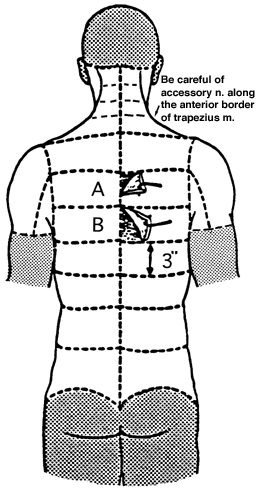
External occipital protuberance Spinous process and spina prominens 3. Begin to remove the skin from the back as shown in Figure 1. (Play movie)
Skin varies in thickness between regions of the body and individuals. Since we are removing only the skin (epidermis and dermis) in some regions, its thickness must be ascertained. Make an initial longitudinal incision in the mid-line of the back beginning at the base of the skull (external occipital protuberance) and continuing caudally to the tip of the spine (coccyx) as shown in Figure 1. Make a transverse incision at right angles to the first anywhere along its length. Using a toothed forceps, turn back the corner of skin at the intersection of the two incisions and measure the skin thickness (dermis and epidermis) with the scalpel. Using the scalpel and your finger as a depth guide, make transverse incisions through the skin as indicated in Figure 1, approximately 8 cm. (3 inches) apart. The skin of the back is usually the thickest. Use the same procedure of measuring thickness and making incisions through the skin in each region of the body.
Figure 1 Begin in the mid to upper back region. To reflect a flap of skin, begin at a corner where two incisions meet at right angles. Pick up the skin with your toothed forceps and with the scalpel cut the skin from the underlying subcutaneous tissue (superficial fascia). Generally, small and controlled cutting motions provide cleaner results.
Because of its density, the dermis presents considerable resistance to the scalpel, while the subcutaneous tissue, being mostly fat, presents little or no resistance. Continue elevating the skin from the subcutaneous tissue until the length of the scalpel swing becomes too long to control easily. Return to the opposite corner of the skin flap and proceed as indicated above until another corner is produced. Working from the corners is much easier and more accurate than long areas of reflection.
4. Separate skin from subcutaneous connective tissue. (Play movie)
For two strips of skin on either side (e.g. A, B Figure 1) just remove the skin, leaving the subcutaneous tissue with nerves and vessels intact. As the skin is subsequently rolled back in these strips the tip to the scalpel should be pointed at the roll of skin and in contact with the dermis. The motion of the scalpel is used in somewhat of a scraping motion rather than strictly cutting. On the deep surface of the dermis, when removed in the proper plane, a "pigskin" or pitted surface is visible. This effect is due to the presence of subcutaneous papillae projecting into the dermis. Keep this constantly in view.
5. Complete the removal of skin and superficial fascia of the back. (Play movie)
For the other strips, remove skin and subcutaneous tissue together (e.g. remove everything down to the deep, investing fascia) as indicated by Figure 1, as far laterally as the mid-line of the side of the body (mid-axillary line). The back of the neck is difficult because of its curvature. Let the head hang by putting a wooden block under the chest to straighten the neck and make access easier. The highest incision extends from the external occipital protuberance to the mastoid process and then along a line from the mastoid process laterally to the tip of the acromion process. Care must be taken not to cut too deeply along this line as the large spinal accessory nerve crosses the region quite superficially (see later for dissection of this nerve).
After the skin is removed examine it and compare differences in thickness, pigmentation, hair, orifices of glands and the grossly visible epidermis and dermis in various areas. Compare with skin of other bodies. Read the introductory comments in your text on connective tissue, subcutaneous tissue (superficial fascia), and fascial spaces. Examine the subcutaneous tissue, noting composition (fat or connective tissue) and relative proportions of each. Some bodies will have no fat, only connective tissue sheets. Compare characteristics of subcutaneous tissue that distinguish male and female. Compare subcutaneous tissue in the different regions you have exposed. Examine the structure of subcutaneous tissue, noting "skeletal network" of connective tissue supporting fat lobules and membranous planes within subcutaneous tissue.
6. Dissect out an exemplary cutaneous nerve or two and clean the trapezius and latissimus dorsi muscles. (Play movie; View images: N 17, 163, 165, 167, 170, 173, 182, 187, 250, TG 1-12, 1-17, 1-21, 4-11, 4-12)
The cutaneous nerves of the back are bilaterally occurring, segmentally arranged, posterior primary rami of spinal nerves. The structure of spinal nerves themselves will be considered in session 3. They pass from the spine through the deep musculature of the back, pierce the investing (deep) fascia to enter the subcutaneous tissue, traverse it to become more superficial, and enter the skin. Map out the areas of cutaneous innervation supplied by these posterior primary rami. Though they enter the subcutaneous tissue near the spine, the precise point of emergence depends upon the region of the body. This point of emergence is medial and deep to the area of distribution in the skin. The nerves parallel one another (3 to 3 1/2 cm apart), are horizontal in the upper half of the trunk and oblique in the lower half.
In those areas where the subcutaneous tissue was preserved, nerves may be located by incising through the superficial fascia in the mid-line and elevating and separating this layer from the deep fascia. As you continue separating these planes laterally, the nerves can be seen leaving the muscle and entering the superficial fascia. Blunt dissection (using probe and forceps) is necessary here as a scalpel or sharp dissection generally severs most of the nerves before they are seen. Many of the nerves may be accompanied by small arteries and veins; the small veins may be filled with blood, giving them a chocolate color, and are more obvious. These vessels supply the skin and subcutaneous tissue and are not specifically named. Identify an exemplary nerve, and if you have time look for the superior cluneal nerves (from L1,2,3). Trace the nerves laterally as far as possible to determine their manner of branching and area of distribution. Note their shape and longitudinal striations. What are these?
(Click on the
Superior cluneal nerves, from spinal nerves L1,2,3, enter the subcutaneous tissue at the lateral border of the erector spinae muscle mass (deep back muscle) and pass obliquely into the buttocks. They can be located at this point of emergence and as they cross the crest of the ilium, since they lie directly on the bony crest.
After finding a few examples of nerves, the subcutaneous tissue of the back should be completely removed down to the investing fascia using a scalpel in much the same manner as used to remove the skin. Leave examples of the cutaneous nerves for subsequent dissections. As you dissect and reflect the muscles, pull the cutaneous nerves through the muscles.
After the subcutaneous tissue has been removed the musculature can be seen. Deep or investing fascia covers the muscles and hides their details. Distinguish between investing and muscular fascia. What is the function of fascia?
7. Reflect trapezius, identify its blood and nerve supply, and locate the greater occipital nerve. (Play movie; View images: N 33, 127, 174, 177, 178, 179, 420, 421, 427, 429, 430, TG 1-09, 1-12, 1-13, 1-16, 7-14, 7-16, 7-17)
For each muscle examined throughout the course, the following characteristics should be observed: origin, insertion, nerve supply, blood supply (arteries and veins), parts and structural arrangement of the muscle, and its relationships (position in the body and its relation to other structures adjacent to it) and a consideration of its action. The focus will be on the action of muscle groups unless a muscle has a unique action. This listing is given in our initial dissection of muscle but is applicable to all subsequent muscles wherever encountered in the body. As you dissect verify your observations with textual descriptions for completeness and accuracy.
HOW TO CLEAN A MUSCLE. The superficial muscles of the entire body are covered with an investing fascia that is relatively opaque and firmly adherent to the muscle. It is continuous with investing fascias of adjacent muscles. Cleaning a muscle requires removal of this fascia and any other connective tissue or fat which may be obscuring the muscle. The fascia is elevated with toothed thumb forceps and then cut away with the scalpel. Remove the fascia by cutting it from the muscle at right angles to the muscle fascicles. Cleaning makes apparent the basic features of a muscle: borders, origin, insertion, and fiber direction. Cleaning does not imply beauty or perfection but only the ability to see the above characteristics.
Origin, insertion, borders, and muscle structure must be visible and verified before the muscle is reflected. To reflect a muscle means to cut one attachment of the muscle and fold it back on itself. Reflection or removal may be done by severing the origin, the insertion, or by cutting through the belly of the muscle as specified.
The muscular fascia on the deep side of a muscle is usually transparent, thin, and easily removed; however, between some muscles there may be additional quantities of loose fascia, fat and connective tissue which impede visibility and have to be removed. What is the function of this fascia?
On the deep surface of a muscle, near the junction of its proximal and middle thirds, is usually the entrance of the artery, vein, and nerve (neurovascular bundle). This is the motor point or hilum. These are usually surrounded by fat and connective tissue and need care in cleaning. There are two methods of locating these important structures. (1) Carefully remove the muscular fascia on the deep side of the muscle until tributaries of the artery, vein or nerve are visible, and then trace these back to the main trunk. (2) Locate the approximate motor point or hilum and probe through the fat until the main trunks are found. In either method only the probe should be used initially.
The arteries are beige. They maintain a cylindrical appearance because of their thick muscular wall. The veins are usually larger than the arteries and are often dark in appearance due to coagulated blood retained within them. They are thin-walled, tortuous and usually paired or plexiform in arrangement. If empty they appear flat or readily collapsible. Nerves appear striped, and in their normal state are fusiform in cross section. The nerve, artery, and vein usually accompany one another to a muscle as a triad, the neurovascular bundle. Where you find one you usually find the others. Expose completely the course of the veins, arteries, and nerves, and observe their particular manner of branching. Why do nerves and vessels (arteries and veins) branch in different manners?
In your dissections, deep veins may be removed after they are cleaned and identified, since they parallel arteries, usually share the same name as the artery, and often obscure the artery and nerve. Do not cut or remove arteries or nerves.
Cleaning is complete when the source, course, and distribution of the artery, vein, and nerve are visible. Since these three characteristics identify arteries, veins and nerves, it is essential that they be seen.
The muscles examined in this dissection are primarily muscles of the pectoral girdle (muscles which attach the upper limb to the trunk, rather than act as prime movers of the upper limb).
For each muscle identify its points of attachment (origin and insertion) on the skeleton so that you may readily become acquainted with the specific bones, their parts and processes as you progress through the dissections. Knowledge of the attachments and where a muscle crosses a joint allows one to predict its action.
Trapezius muscle. Clean and define origin and insertion and borders of this muscle. Determine arrangements of muscle fibers, divisions of the muscle, kinds of attachments (tendon, aponeurosis, muscle). Define the superior border (between superior nuchal line of the occipital bone and clavicle) but do not cut deeply along this border as the accessory nerve is superficial there. Reflect the muscle by incising along its origin. To do this, pick up the muscle and separate it from underlying muscle before cutting. Progress in this fashion from T12 upward until you have completely severed its origin. In the thoracic region the muscle takes origin from the spines of the vertebrae and intervening ligaments. In the cervical region the spines are short and the muscle takes origin from a strong ligament, the ligamentum nuchae. Lift the trapezius up off of the underlying neck muscles. Cut the cranial attachment of this muscle, inferior to (and away from) the superior nuchal line.
It will also be necessary to sever the muscle along its insertion into the spine of the scapula and along the medial border of the acromion. Do not sever its clavicular attachment. In reflecting use only the probe to separate it from deeper structures in order to avoid cutting arteries and nerves. It will be necessary to cut the muscular perforating branch of the dorsal scapular artery and vein to reflect the lower part of the muscle. This artery arises from the dorsal scapular artery (to be seen later). It enters the lower third of the trapezius muscle after penetrating between rhomboideus major and minor or through rhomboideus major.
Identify and trace the subtrapezial plexus of nerves. Distinguish between the accessory nerve (Cranial Nerve XI) (reaching the muscle from the posterior triangle of the neck along the deep surface of the superior border of the muscle) and branches of cervical nerves 3 and 4 (C3, 4) reaching the muscle from a position anterior and deep to the levator scapulae muscle. These nerves are branches of anterior primary rami of spinal nerves. Where do they combine to form the subtrapezial plexus?
As you reflect the trapezius near the occipital bone, stay close to its under surface. The greater occipital nerve is from the dorsal primary ramus of C2. It will lie on the underlying muscle mass about 2cm below and 2 to 3cm lateral to the external occipital protuberance. Try to locate it (you may need your instructor's help with this one). It is a large nerve extending vertically into the scalp and can be recognized by its association with the large occipital artery and vein.
8. Reflect latissimus dorsi, levator scapulae, and the rhomboids and identify their blood and nerve supply. ( Play movie; View images: N 33, 174, 421, 424, 427, 429, 430, 477, TG 1-12, 1-13, 2-06A, 2-06B, 2-07, 2-09, 2-13, 2-15, 7-15)
Latissimus dorsi muscle. Define origin and borders of muscle. What is the thoracolumbar fascia (aponeurosis)?
Levator scapulae, rhomboideus minor, and rhomboideus major muscles insert along the medial border of the scapula in serial order. Beginning at the medial border of the scapula, trace each muscle to its origin. Note and separate the slips or heads of origin of the levator scapulae m. passing to transverse processes of cervical vertebrae. The rhomboideus minor and rhomboideus major muscles may not be separate; they are often referred to collectively as the Rhomboids. Branches of the same nerves (cervical 3 and 4) that supply trapezius supply the levator scapulae. Using the branches of C3 and C4 that you have identified, follow their proximal course along the anterior border of the levator scapulae and identify them as they enter the muscle. Cut the origins of the rhomboideus minor and major and reflect them. Near their insertion along the medial border of the scapula (well hidden by fat and connective tissue), locate the dorsal scapular nerve (C5) and dorsal scapular artery and vein. Trace the nerve and vessels to the rhomboid muscles. Look for the dorsal scapular nerve sending fibers to the lower part of the levator scapulae muscle. You may notice thin, aponeurotic muscles under the rhomboids above and the latissimus dorsi below. These are the serratus posterior superior and inferior muscles. They are frequently reflected with the rhomboids and or latissimus dorsi. They are accessory respiratory muscles and of minor importance.