|
|
|
||||||||||||
Dissector Answers - Pectoral Region & Breast |
|||||||||||||
Learning Objectives:
Upon completion of this session, the student will be able to:
- Describe the general gross features of the breast and its blood supply.
- Describe the lymphatic drainage of the breast.
- Identify the muscles of the pectoral region, their related fascia, nerve and regional blood supply, and general functions.
- Describe the general features of the circulatory system.
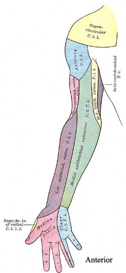
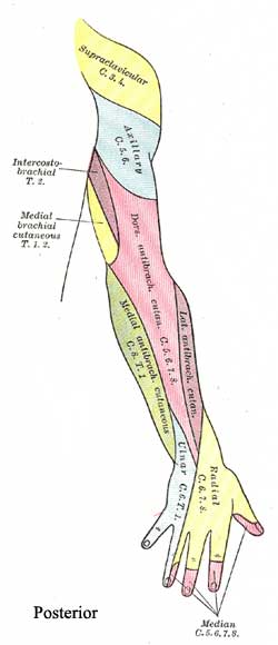
- Identify and demonstrate the areas of distribution of the major cutaneous nerves of the upper limb.
- Identify and demonstrate the major superficial veins of the upper limb.
Learning Objectives and Explanations:
1. Describe the general gross features of the breast and its blood supply. (W&B 107-110, N 182A, 182B, TG 2-10A, 2-10B, 2-11A)The mammary gland is a modified sweat gland. It is entirely contained within subcutaneous tissue. The most important internal gross features are glandular, namely secretory glands, lactiferous ducts, and lactiferous sinuses. (It is the glandular nature of the breast that makes it a common site for the development of cancer.) Externally, an important feature is the nipple, which is surrounded by the areola. Each of the approximately 20 lactiferous sinuses have an individual opening on the nipple.2. Describe the lymphatic drainage of the breast. (W&B 109, N 184, image, TG 2-11B)
A breast's arterial supply is derived from branches of the internal thoracic artery (including anterior intercostals), the lateral thoracic artery, the thoracoacromial artery, and posterior intercostal arteries. Venous drainage follows arterial supply, primarily draining into the axillary vein, but also draining some blood into the internal thoracic vein.Lymph passes from the nipple, areola, and lobules to the subareolar lymphatic plexus. From there:3. Identify the muscles of the pectoral region, their related fascia, nerve and blood supply, and general functions. (W&B 111-113, N 188, 189, TG 2-12A, 2-12B)Lymphatic vessels in the skin of the breast drain into the axillary, inferior deep cervical, infraclavicular, and parasternal lymph nodes.
- MOST (75%) of the lymph goes to the axillary lymph nodes, via the pectoral lymph nodes. (It is extremely important to consider the axillary nodes when performing a breast exam on a patient.)
- Most of the rest goes to the parasternal lymph nodes.
- A small amount of lymph goes to the opposite breast.
- A small amount of lymph goes to the abdominal wall and downward.
Lymph from the axillary lymph nodes subsequently drains into the subclavian lymph trunk. Lymph from parasternal nodes enters the bronchomediastinal trunk.4. Describe the general features of the circulatory system. (W&B 17-24)
Muscle Origin Insertion Action Innervation Artery Notes pectoralis major medial 1/2 of the clavicle, manubrium & body of sternum, costal cartilages of ribs 2-6, sometimes from the rectus sheath of the upper abdominal wall crest of the greater tubercle of the humerus flexes and adducts the arm, medially rotates the arm medial and lateral pectoral nerves (C5-T1) pectoral branch of the thoracoacromial trunk the deep fascia on its anterior surface should not be fused to the fascia of the mammary gland - if it is, this is an important clinical sign indicating breast disease pectoralis minor ribs 3-5 coracoid process of the scapula draws the scapula forward, medialward, and downward medial pectoral nerve (C8, T1) pectoral branch of the thoracoacromial trunk branches of medial pectoral nerve usually pierce pectoralis minor to reach the pectoralis major muscle serratus anterior ribs 1-8 or 9 medial border of the scapula on its costal (deep) surface it draws the scapula forward; the inferior fibers rotate the scapula superiorly long thoracic nerve (from ventral rami C5-C7) lateral thoracic a. a lesion of long thoracic nerve will cause winging of the scapula (i.e., the medial border of the scapula falls away from the posterior chest wall and looks like an angel's wing) (Latin, serratus = to saw)
The human circulatory system actually contains two complete "circuits". The right side of the heart receives deoxygenated blood from the body and pumps it to the pulmonary circuit (i.e., the lungs). The left side of the heart receives oxygenated blood from the lungs and pumps it to the rest of the body.5. Identify and demonstrate the areas of distribution of the major cutaneous nerves of the upper limb. (W&B 102-106, N 429,430,431, 432,474,477, 478, 479, 480, 481, 482, TG 2-02,2-14,2-51A,2-51B,2-52A,2-52B,2-33)
The general order of vessels, beginning with the aorta, is artery, arteriole, capillary, venule, vein, then back into the heart. There are exceptions - the hepatic portal system is the major one you will deal with soon. In gross anatomy, you will only be concerned with structures that are visible without a microscope, i.e. arteries and veins.6. Identify and demonstrate the major superficial veins of the upper limb. (W&B 101-103, N 189,479, 480,483, TG 2-02,2-53)
Images from "Anatomy of the Human Body" by Henry Gray are provided by:
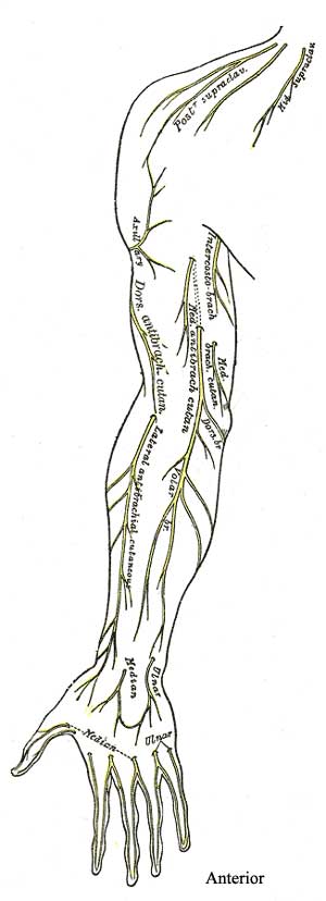
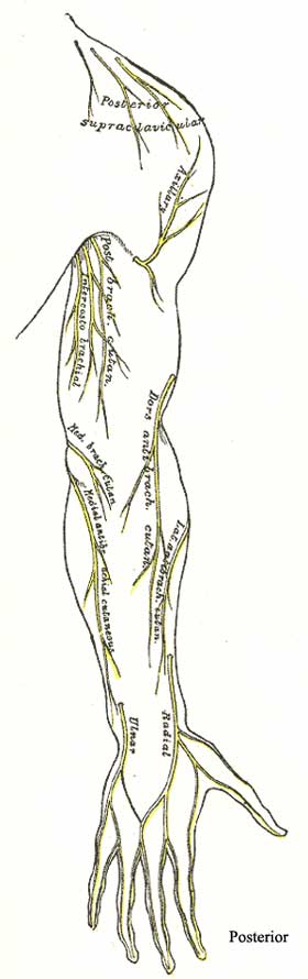
The major cutaneous nerves of the upper limb:
- radial nerve:
- posterior brachial cutaneous nerve
- posterior antebrachial cutaneous nerve
- inferior lateral brachial cutaneous nerve
- musculocutaneous nerve (continues below the elbow as the lateral antebrachial cutaneous nerve)
- medial cord of the brachial plexus:
- medial brachial cutaneous nerve
- medial antebrachial cutaneous nerve
- Also remember that the ulnar and radial nerves both have superficial extensions which cover the dorsum of the hand, namely the dorsal cutaneous branch of the ulnar nerve and the superficial branch of radial nerve.
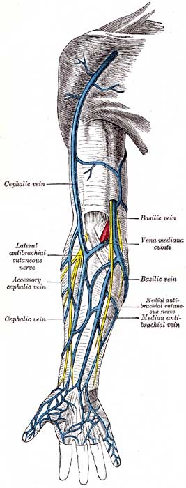
Arising from the dorsal venous network (arch) on the dorsum of the hand are the two most important superficial veins: the cephalic and basilic veins. The cephalic vein ascends along the lateral aspect of the forearm. (This side of your arm is considered "cephalic" because the limbs develop projecting laterally from the body, with the lateral side closest to the head. Or, you can remember that the cephalic side is on the side of your thumb, the digit you stick in your cephalic orifice (mouth) when you are a baby.) Upon completing its ascent, the cephalic vein crosses through the deltopectoral groove before diving deep through the fascia and joining the axillary vein. The basilic vein ascends on the medial aspect of forearm and pierces the deep fascia of the inferior arm. It ascends to unite with the paired brachial veins, forming the axillary vein. The cephalic and basilic veins are connected by the median cubital vein in the cubital fossa.
Cultural enrichment: Check out these sections from the 1918 version of Gray's Anatomy of the Human Body! Some of the terms are (of course) out-of-date, but the illustrations are timeless. The Sternum - The Muscles Connecting the Upper Extremity to the Anterior and Lateral Thoracic Walls - The Mammae (Breasts) - The Lymphatics of the Upper Extremity - Surface Anatomy of the Thorax - Surface Markings of the Thorax
Questions and Answers:
7. From where do the lateral cutaneous branches (anterior and posterior) of intercostal nerves arise and to where do they distribute?The lateral cutaneous branches of a typical intercostal nerve arise lateral to the angles of the ribs, divide into anterior and posterior branches, and supply the skin of the thoracic and abdominal walls. (W&B 108, N 188, 189, TG TG4-02, TG4-11)8. Is any pinkish mammary glandular tissue visible?It is likely that your cadaver does not have any appreciable mammary tissue. Most of the cadavers are elderly.9. What are suspensory ligaments of the mammary glands?The suspensory ligaments are fibrous condensations of the connective tissue stroma which are prominent in the superior part of the mammary gland. These help support the lobules of the gland and attach it to the dermis of the overlying skin. (N 182A, 182B, TG 2-10A, 2-10B)10. Consider blood supply, nerve supply, and lymphatic drainage of the breasts.Blood: A breast's arterial supply is derived from branches of the internal thoracic artery (including anterior intercostals), the lateral thoracic artery, the thoracoacromial artery, and posterior intercostal arteries. Venous drainage follows arterial supply, primarily draining into the axillary vein, but also draining some blood into the internal thoracic vein.11. Why are lateral pectoral and medial pectoral nerves reversed (with respect to their names and relative locations) from what you would expect?
Nerves: The skin of the breast is innervated by anterior and lateral cutaneous branches of the 2nd through 6th intercostal nerves. For example, the area around the nipple and areola is innervated via the T4 spinal nerve.
Lymphatic drainage (W&B 109, N 184, TG 2-11B, image): Lymph passes from the nipple, areola, and lobules to the subareolar lymphatic plexus. From there:Lymphatic vessels in the skin of the breast drain into the axillary, inferior deep cervical, infraclavicular, and parasternal lymph nodes.
- MOST (75%) of the lymph goes to the axillary lymph nodes, via the pectoral lymph nodes. (It is extremely important to consider the axillary nodes when performing a breast exam on a patient.)
- Most of the rest goes to the parasternal lymph nodes.
- A small amount of lymph goes to the opposite breast.
- A small amount of lymph goes to the abdominal wall and downward.
Lymph from the axillary lymph nodes subsequently drains into the subclavian lymph trunk. Lymph from parasternal nodes enters the bronchomediastinal trunk.Both of these nerves supply the pectoralis major. The "medial" and "lateral" designations of these two nerves refers to the cords of the brachial plexus from which they are derived. (Do not worry about this now, but it will haunt you later!) They are not topographic designations. (To remember which is which, think about the Medial pectoral nerve being a Major nerve which supplies both the pectoralis Major and the pectoralis Minor. The Lateral nerve is a Little nerve, and only innervates pectoralis major.) (N 430, TG 2-12B, 2-13)12. Where does the cephalic vein terminate?The cephalic vein passes between the deltoid and pectoralis major muscles and empties into the termination of the axillary vein. (Latin/Greek, cephalicus/kephalikos = head) (N 424, TG 2-12A, 2-02AB)13. Identify the dorsal digital veins and intercapitular veins. What do these do?Dorsal digital veins drain the dorsal cutaneous aspect of the fingers. There are two per finger. Intercapitular veins are found between the knuckles (heads of the metacarpals). They drain the fingers and palm to the dorsal side of the hand. N480,TG2-0214. Does the basilic vein perforate the brachial fascia in your cadaver? As you traced the basilic vein did you find accompanying nerves? What are these?The basilic vein normally perforates the brachial fascia above the medial epicondyle, or even as high as mid-arm. Distally near the hand, the vein is accompanied by the dorsal branch of the ulnar nerve. As the vein ascends it is accompanied by the anterior and posterior branches of the medial antebrachial cutaneous nerve. N479,TG2-0215. Where does the cephalic vein penetrate the deep fascia?As mentioned above, the cephalic vein penetrates the deep fascia after it has traversed the deltopectoral groove at the clavicle. N424,TG2-0716. Does the cephalic vein extend into the arm?Of course, though in some arms it may be very small. N479,N480,TG2-0217. What is the course and direction of the median cubital vein? Look at other arms to determine pattern. What are other variations?Median cubital vein shunts blood from cephalic vein obliquely across the cubital fossa to reach the basilic vein. There is often a median antebrachial vein lying on the anterior forearm that splits to drain to both cephalic and basilic veins at the cubital fossa.18. Trace the superficial branch of the radial nerve into the hand and identify its dorsal digital branches and their areas of distributions. Do any branches communicate with branches of the ulnar nerve?The two nerves usually communicate on the dorsum of the hand. N480,N481,TG2-02,TG2-51