 |
|||||
|
|
|
||||||||||||
Dissector Answers - Peritoneal Cavity & Intestines |
|||||||||||||
Learning Objectives:
Upon completion of this session, the student will be able to:
- Describe the basic organization of the peritoneum and peritoneal cavity, including subdivions, mesenteries, and ligaments.
- Describe the basic anatomy of the large and small intestines, including blood supply and internal structure.
- Know the pattern of diaphragmatic musculature and its fasciae, and its functional significance in respiration.
- Know the three major passageways through the diaphragm and the structures traversing them.
- Describe the position and vertebral level for all branches of the abdominal aorta and the inferior vena cava, and the reason for the difference in their patterns.
- Identify the thoracic and lumbar splanchnic nerves and the collateral ganglia or regional subdivisions of the preaortic plexus to which each functionally relates.
- Recall the concept of perivascular plexuses, their position, nomenclature, and nerve fiber components.
- Describe the parasympathetic innervation of the GI tract.
- Identify the cisterna chyli and describe the general pattern of lymphatic drainage to the thoracic duct.
- Identify and demonstrate the abdominal attachments of the two major posterior abdominal wall muscles and know the action of these muscles upon the vertebral column.
- Locate the lumbar sympathetic trunk and white and gray rami communicantes; explain the reason for the inferior limit of the white rami.
Learning Objectives and Explanations:
1. Describe the basic organization of the peritoneum and peritoneal cavity, including subdivisions, mesenteries, and ligaments. (W&B 436-446, N 264,267,273,276,335,337, TG 5-12A, 5-12B, 5-13, 5-14, 5-15, 5-16B, 5-16C, 5-29, 5-31, 5-42, 5-43)2. Describe the basic anatomy of the large and small intestines, including blood supply and internal structure. (W&B 477-488, N 291,295,296,300,301,302, TG 5-12A, 5-12B, 5-13, 5-14, 5-15, 5-16B, 5-16C, 5-29, 5-43)The peritoneum, like the pericardium and pleura, is a serous membrane that invests viscera. It is comprised of parietal and visceral peritoneum. There are many specializations of the peritoneum. All of the special structures that will be covered here are composed of two layers of peritoneum (much like the pulmonary ligament). They differ in location and what they connect. (Greek, peritonaion = stretch around)
Mesenteries: result from the invagination of "intraperitoneal" organs into the sac. The mesenteries connect viscera to the posterior abdominal wall and are VERY important in that they conduct blood vessels and nerves. (There are no vessels within the peritoneal cavity, of course.) The mesentery of the colon is usually called the "mesocolon". For example, we speak of the "transverse mesocolon" and the "sigmoid mesocolon". (The other parts of the colon are not completely invested by peritoneum, and are therefore "retroperitoneal".) Also, often "the mesentery" refers specifically to the mesentery of the small intestine. (Greek, mes = in the midst of, enteron = intestine)
Omenta: generally refers to a free fold of peritoneum. This is exemplified by the greater omentum, which attaches to the stomach, droops far down into the abdominal cavity, and comes back up to attach to the transverse colon. The lesser omentum, on the other hand, is not really "free". It connects the stomach to the liver, and its membranous portion is called the hepatogastric ligament.
Ligaments: connect organs to one another or to the abdominal wall. Usually, in this case, we are referring to the stomach or the liver.
- falciform ligament: between liver and abdominal wall
- hepatogastric ligament: between stomach and liver, part of lesser omentum
- hepatoduodenal ligament: liver and duodenum, part of lesser omentum
- gastrophrenic ligament: stomach and diaphragm
- gastrosplenic ligament: stomach and spleen
- gastrocolic ligament: stomach and transverse colon
- splenorenal ligament: between the spleen and the left kidney
There are many folds and fossae that can be described in the peritoneum. Some have already been discussed (see Abdominal Wall Dissector Answers). Others are inconstant and not extremely important (see W&B 445-446). A few structures should be noted, due to their clinical importance. (Appendectomy anyone?) They are found in the vicinity of the cecum, are listed here superior to inferior:
- anterior cecal fold (constant): connects "the mesentery" to anterior cecum. It contains the anterior cecal artery.
- superior ileocecal fossa (constant): between the anterior cecal fold and ileocecal junction.
- ileocecal fold (variable): connects terminal ileum to cecum. It crosses the root of the vermiform appendix and is avascular. (Latin, vermiform appendix is literally "worm-shaped appendage")
- inferior ileocecal fossa (variable): between ileocecal fold and mesoappendix.
- mesoappendix: a continuation of "the mesentery" that carries the blood supply for the appendix - the appendicular artery.
The peritoneal cavity, analogous to the pleural cavity, is a space between the two layers of peritoneum. Fluid in this space provides lubrication, allowing the abdominal viscera to move around. The peritoneal cavity contains no viscera. There are two major subdivisions of the peritoneal cavity:
Greater sac: the main part of the peritoneal cavity. It is divided into two sections. The supracolic and infracolic compartments are divided by the transverse mesocolon. The infracolic compartment is further divided into right and left infracolic spaces by "the mesentery".
Lesser sac (omental bursa): lies posterior to the stomach. It is divided into two sections, a superior and an inferior recess.
The passage between the greater and lesser sacs is the omental (epiploic) foramen, posterior to the hepatoduodenal ligament.
3. Know the pattern of diaphragmatic musculature and its fasciae, and its functional significance in respiration. (W&B 497-499, N 194,195,263 or TG5-33)The small intestine comprises the duodenum, jejunum, and ileum. The first and shortest portion of the small intestine is the duodenum, but due to its intimate relationship with the liver and pancreas, it will be considered with those structures. The remainder of the small intestine is divided into the jejunum and the ileum. The border between them is not distinct, but the jejunum is usually thought to lie mostly in the left upper quadrant, comprising about 40% of the total length, while the ileum lies mostly in the right lower quadrant, providing the remaining 60%.
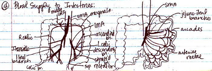
The small intestine contains circular folds of tissue that are covered with villi. (This will become important in histology.) Added to the fact that the long tube is coiled in the abdominal cavity, there is A LOT of surface area over which nutrients can be absorbed. It is completely supplied by the superior mesenteric artery (SMA), via 15 or so intestinal (jejunal and ileal) arteries. These smaller branches eventually turn into arterial arches (arcades), which resemble a colonnade, providing collateral circulation to different sections of the organ. Straight arteries (vasa recta) then jump from the arcades to the tissue.
The colon is considered in five sections. From the cecum, where the small intestine attaches, the ascending colon travels up to the right colic (hepatic) flexure. From there, the transverse colon moves across to the left colic (splenic) flexure. Then, the descending colon travels down, becoming the sigmoid colon which ends in the rectum.
The colon, or large intestine, has three features that distinguish it from the small intestine:
- Its longitudinal muscle forms three teniae coli, instead of being continuous all the way around.
- The teniae coli are actually shorter than the colon, so the large intestine forms haustra coli (bulges, sacculations).
- The surface of the colon has fat-filled tabs, called omental appendages (appendix epiploica).
Note: The teniae coli come together at the base of the appendix, which can help in locating the appendix on the cecum.The colon receives blood from both the superior mesenteric artery and the inferior mesenteric artery (IMA). The SMA supplies the colon up to the splenic flexure, via the following branches:
- ileocolic artery: divides into ileal and colic branches to supply the cecum and the ascending colon. The ileocolic artery also gives off the appendicular artery that supplies the vermiform appendix.
- right colic artery: supplies the ascending colon
- middle colic artery: supplies the transverse colon
The IMA supplies the rest of the colon via:
- left colic artery: supplies the descending colon
- sigmoid arteries: 3 or 4 branches that supply the descending and sigmoid colon
- superior rectal artery: supplies upper rectum
If you draw pretty pictures like this on occasion, especially when connectivity is important, you'll be a happy medical student! Thanks to Susana Gonzalez!
4. Know the three major passageways through the diaphragm and the structures traversing them. (W&B 499, N 194,195,263 or TG 5-33)
This view of the diaphragm shows all of the structures from which the muscle fibers take origin. The diaphragm is divided into three parts on the basis of these muscle fiber origins:
- sternal part: small origin of muscle on the posterior aspect of the xiphoid process
- costal part: origins from the cartilage and bone of the lower six ribs
- lumbar part: all of the rest, including origins from the bodies of the first three lumbar vertebrae (forming two crurae) and from the arcuate ligaments (labeled "lumbocostal arches" above).
Think of the diaphragm as a round dome tent. All of the fibers originate from the ground, where the tent is staked down. The stakes in front are the xyphoid process, those in back are the lumbar vertebrae. The posterolateral stakes are the arcuate ligaments, while the anterolateral stakes are the ribs. They all insert into the central tendon, which is the top of the tent where everything comes together.
Some parts of the diaphragm are:
- right crus: takes origin from L1-L3. It splits to enclose the esophagus, so the esophageal hiatus is (usually) entirely formed by the right crus. Fibers from the right crus intermingle with the fibers from the left crus at the aortic hiatus. (Latin, crus = resembling leg or legs)
- left crus: takes origin from L1-L2. It is smaller and shorter than the right crus. It sometimes contributes something to the esophageal hiatus. Fibers from the left crus intermingle with the fibers from the right crus at the aortic hiatus. (Latin, crus = resembling leg or legs)
- medial arcuate ligaments: thickening of psoas major fascia. Fibers taking origin from here, along with those from the lateral arcuate ligament, fill in the "gap" between the crura and the costal part of the diaphragm. They are labeled "medial lumbocostal arches" above. (Latin, arcuare = to bend like a bow)
- lateral arcuate ligaments: thickening of the quadratus lumborum fascia. Fibers taking origin from here, along with those from the medial arcuate ligament, fill in the "gap" between the crura and the costal part of the diaphragm. They are labeled "lateral lumbocostal arches" above. (Latin, arcuare = to bend like a bow)
The diaphragm is the primary muscle of respiration. Contraction of the fibers pulls the central tendon inferiorly, increasing the volume (and decreasing the pressure) of the thoracic cavity. Since good ole PV=nRT creates a pressure gradient between the inside and the outside, air rushes in to compensate. Secondary to this, the volume of the abdominal cavity is decreased, raising its pressure. Sometimes you do actually want to do this, like during defecation or parturition. The diaphragm can assist the abdominal wall muscles here.
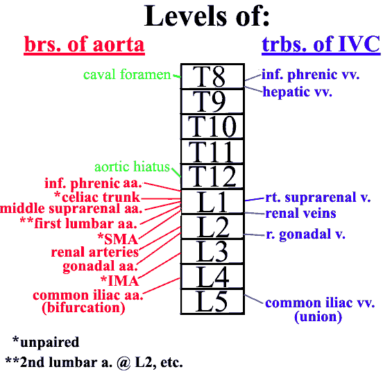
5. Describe the position and vertebral level for all branches of the abdominal aorta and the inferior vena cava, and the reason for the difference in their patterns. (W&B 507-512, N 264, 265 or TG 5-34, 5-34)
- vena caval foramen: T8 - transmits the IVC, right phrenic nerve branches, and small lymphatic vessels
- esophageal hiatus: T10 - transmits the esophagus, anterior and posterior vagal trunks, branches of left gastric vessels, and small lymphatic vessels (Latin, hiare = to yawn... referring to an interruption)
- aortic hiatus: T12 (not really a hole in the diaphragm, but more of a dent in its edge) - transmits descending aorta, thoracic duct, and sometimes the azygous vein. (Latin, hiare = to yawn... referring to an interruption)
6. Identify the thoracic and lumbar splanchnic nerves and the collateral ganglia or regional subdivisions of the preaortic plexus to which each functionally relates. (W&B 503, N 209,318 or TG 4-38, 5-39)
The tributaries of the inferior vena cava, in part, follow the branches of the abdominal aorta. There are some differences, however. The major one is that the SMV and the IMV are part of the portal system, and therefore have nothing to do with the IVC. Also, the left gonadal veins drain into the left renal vein, instead of directly into the IVC.
7. Recall the concept of perivascular plexuses, their position, nomenclature, and nerve fiber components. (N 318, 319, 322, 323 or TG 5-39,8-16,8-17)The presynaptic (preganglionic) fibers of the thoracic splanchnic nerves originate from the sympathetic trunk in the thorax and run through the diaphragm to provide most of the sympathetic innervation to abdominal viscera. The greater thoracic splanchnic nerve comes from T5-T9 and synapses in the celiac ganglion. The lesser thoracic splanchnic nerve comes from T10-11 and synapses in the aorticorenal ganglion. The least thoracic splanchnic nerve goes from T12 to the renal plexus.
The presynaptic (preganglionic) fibers of the lumbar splanchnic nerves originate from the sympathetic trunk in the abdomen. They provide small fibers to all of the preaortic plexuses, including the celiac, superior mesenteric, intermesenteric, and superior hypogastric plexuses.
The point of all of this is to shut down the GI tract when energy needs to be diverted to other activities. In addition, fibers go to the smooth muscle in the walls of arteries and arterioles to regulate blood flow and blood pressure.
8. Describe the parasympathetic innervation of the GI tract. (W&B 456-457 (stomach), 465-466 (duodenum), 482 (SI), 488 (LI))Perivascular plexuses are just that - plexuses of autonomic nerves that run along vessels in order to reach their targets. Sometimes they are using the vessel as a convenient roadway to reach a visceral target. In other cases, since blood vessel smooth muscle receives sympathetic innervation, the vessels are the target. They are named according to the vessel upon which they lie.
This is not an enormously important topic, but is one that is worth thinking about. You will see it again, especially while studying the head and neck.
9. Identify the cisterna chyli and describe the general pattern of lymphatic drainage to the thoracic duct. (W&B 412, 501, TG 5-37)We saw the same question in the session on the stomach. Below is the same answer.
Parasympathetic innervation to most of the GI tract, from the stomach to the splenic flexure of the colon, is originally from the vagus nerves (CN X) (N 228,309,310,311,314A,314B or TG 4-37,8-16,8-17). Along the way they will form various plexuses and then reorganize into nerves, but it is all vagus derived. (For example, after the esophageal plexus, fibers come back together to form the anterior and posterior vagal trunks, which supply the stomach and other viscera.) For the portions of the colon distal to the splenic flexure, the parasympathetic innervation is from S2-S4, via the pelvic splanchnic nerves and the inferior hypogastric plexus. (Recall that arterial blood supply to the colon also had a "transition" near the splenic flexure.)
In addition to this information from the previous lab, there is one small point. The parasympathetic fibers coming from the vagus nerve, innervating the GI tract from the stomach through the proximal 2/3 of the transverse colon, follow some of the same paths as the sympathetics from the thoracic splanchnic nerves. So, for example, they will pass through, but not synapse within, the celiac ganglion. Parasympathetic nerves usually synapse within the target tissue.
On the other hand, the parasympathetic fibers that innervate the distal portion of the colon come from S2-S4 via the pelvic splanchnics. They do not follow along the same paths as the sympathetic fibers, which, in this case, go through the inferior mesenteric plexus. Branches of the pelvic splanchnic nerves reach the hindgut by passing up over the left pelvic brim and through the fusion fascia to reach the splenic flexure, and the descending and sigmoid colon. Of course, pelvic splanchnic nerves also distribute to pelvic viscera, such as the rectum, but we can save that story for the pelvis.
10. Identify and demonstrate the abdominal attachments of the two major posterior abdominal wall muscles and know the action of these muscles upon the vertebral column. (W&B 513-515, N 263 or TG 5-33)In 25-50% of cases, the inferior portion of the thoracic duct includes a dilated portion called the cisterna chyli (chyle cistern). When present, all of the lymph trunks draining the abdomen and lower limbs dump into it, as well as the most inferior intercostal lymph trunks. When it is not present, these trunks simply empty into the thoracic duct.
See the pages indicated above for good diagrams of the general pattern of lymphatic drainage of the body. Especially important in the abdomen is the intestinal lymph trunk, which carries all of the fat from those double cheeseburger combo meals from the small intestine to the thoracic duct.
For more than you ever wanted to know about anatomical variation, the University of Iowa has a great site, an "Illustrated Encyclopedia of Human Anatomic Variation". Here is a quick and dirty link to the section on the thoracic duct and cisterna chyli.
The anatomy tables pretty much say it all:11. Locate the lumbar sympathetic trunk and white and gray rami communicantes; explain the reason for the inferior limit of the white rami. (W&B 503, N N160,N267 or TG 5-38,5-39,5-40,5-41)
Muscle Origin Insertion Action Notes iliacus iliac fossa and iliac crest; ala of sacrum lesser trochanter of the femur flexes the thigh; if the thigh is fixed it flexes the pelvis on the thigh inserts in company with the psoas major m. via the iliopsoas tendon iliopsoas iliac fossa; bodies and transverse processes of lumbar vertebrae lesser trochanter of the femur flexes the thigh; flexes and laterally bends the lumbar vertebral column a combination of the iliacus and psoas major mm. psoas major bodies and transverse processes of lumbar vertebrae lesser trochanter of femur (with iliacus) via iliopsoas tendon flexes the thigh; flexes & laterally bends the lumbar vertebral column the genitofemoral nerve pierces the anterior surface of the psoas major m. psoas minor bodies of the T12 & L1 vertebrae iliopubic eminence at the line of junction of the ilium and the superior pubic ramus flexes & laterally bends the lumbar vertebral column absent in 40% of cases quadratus lumborum posterior part of the iliac crest and the iliolumbar ligament transverse processes of lumbar vertebrae 1-4 and the 12th rib laterally bends the trunk, fixes the 12th rib the lateral arcuate ligament of the diaphragm crosses the anterior surface of the quadratus lumborum m. 12. Describe the four common locations of porto-caval anastomosis. (N320 or TG 5-28)The white rami communicantes are only found as low as the L2 level. Inferior to that there are no sympathetic neuron cell bodies in the spinal cord, and are therefore no sympathetic fibers emerging from the cord into the spinal nerves. Since the white rami communicantes conduct fibers from the spinal nerve to the ganglia, they are unnecessary below L2. But, there are sympathetic fibers in the chain that have traveled from points superior, so there are gray rami communicantes, conducting fibers from the ganglia back onto the spinal nerve, along the chain's entire length.
There are four sites of porto-caval anastomosis described: distal esophageal veins, rectal venous plexus, paraumbilical veins, and posterior abdominal wall veins. These usually appear in roughly this order when portal hypertension, usually due to liver cirrhosis, causes pressure within the portal veins to increase because it has difficulty in passing through the liver sinusoids. The portal venous blood then finds alternate routes back into the caval venous system, bypassing the liver. In rough order of appearance in the progression of this condition:
distal esophageal veins: the thoracic esophagus drains to the azygos system of veins in the chest, while tributaries to the left gastric vein drain the distal esophagus ultimately into the portal vein. Retrograde passage of blood from the portal vein into these distal esophageal veins causes esophageal varicies, enlarged veins within the walls of the esophagus. If these rupture, the blood will be digested and produce black feces.
rectal venous plexus: the superior rectal vein drains the rectum into the inferior mesenteric vein and ultimately the portal vein, while the distal rectum and anal canal drain into the middle and inferior rectal veins, which drain into the internal iliac vein and ultimately into the inferior vena cava. It is commonly thought that retrograde flow of blood down the superior rectal tributaries engorge the veins in the anal columns and cause internal hemorrhoids. However, it has recently been shown that it is more accurate to describe these enlarged veins as within the rectal venous plexus within the walls of the rectum.
paraumbilical veins: very small veins called paraumbilical veins lie within the falciform ligament and drain into the portal vein within the liver. The paraumbilical veins have connections to veins draining the anterior abdominal wall, and retrograde blood in the paraumbilical veins will enlarge the anterior abdominal wall veins. The superficial veins of the anterior abdominal wall will be most visible, of course, and their engorgement, radiating from the umbilicus, is called caput medusa (after Medusa, the woman with the snake hair-do).
posterior abdominal wall veins: given enough back-pressure within the portal venous system, blood enlarges all of the tributaries into the portal system. The portal tributaries of the secondarily retroperitoneal organs (duodenum, pancreas, ascending and descending colon) can form anastomoses with veins of the posterior abdominal wall, the lumbar veins, that ultimately drain into the inferior vena cava usually.
Cultural enrichment: Check out these sections from the 1918 version of Gray's Anatomy of the Human Body! Some of the terms are (of course) out-of-date, but the illustrations are timeless.
The Small Intestine - The Large Intestine - The Abdominal Aorta - Surface Anatomy of the Abdomen -Surface Markings of the Abdomen - The Muscles of the Thorax (diaphragm)
Questions and Answers:
1. What is the vertebral level of an imaginary horizontal line drawn between the right and left iliac crests? (N 248 or TG 5-03)The line between the iliac crests is at the level of L4.2. Differentiate between the abdominal cavity and the peritoneal cavity. (N 335,336,337, TG 5-42A, 5-42B, 5-43A, 5-43B)The peritoneal cavity is within the abdominal cavity, which is continuous with the pelvic cavity. The peritoneal cavity is a potential space, like the pleural and pericardial cavities, between the parietal and visceral layers of the peritoneum. It allows the gut to move with a minimum of friction.3. What is the suspensory muscle of the duodenum? (N 262, TG 5-26)The suspensory ligament of the duodenum (more properly called the suspensory muscle of the duodenum or the Ligament of Treitz) is the structure that suspends the duodenojejunal flexure from the diaphragm. It is continuous with the right crus of the diaphragm.4. Locate the ileocecal junction. At what level is it found? (N 260,273, TG 5-15)The ileocecal junction is just posterior to the anterior superior iliac spine, in the iliac fossa, which is at L4-L5. Another way to say that is that it is right on or just inferior to the transtubercular line, in the right inguinal region.5. Examine "the mesentery" (of the jejunum and ileum), noting its body wall attachment. How long is this attachment? What structures does it cross? (N 263,295, TG 5-13, 5-14A, 5-29)6. Review the development of "the mesentery" from the primitive dorsal mesentery. (Learning Module)The root of the mesentery is approximately 15cm long. It passes inferiorly and to the right, from the duodenojejunal junction to the iliocecal junction, crossing:
- ascending and horizontal parts of the duodenum
- abdominal aorta
- inferior vena cava
- right ureter
- right psoas major muscle
- right testicular or ovarian vessels
7. What is the location of the small intestine within the peritoneal cavity? (N 260,261, TG 5-16A, 5-42A, 5-42B, 5-43A, 5-43B)Initially the entire primordial gut is suspended in the center of the abdominal cavity by a "dorsal mesentery", which is attached to the midline of the posterior body wall. As development progresses, some things start twisting around, some become retroperitoneal, and some do both! When all is said and done, most of the duodenum is retroperitoneal, as is the ascending and descending parts of the colon. Between these two points we have an intraperitoneal structure, the small intestine, that is attached to the body wall via "the mesentery". (That is why the root of "the mesentery" runs from the duodenojejunal junction to the ileocecal junction.) Although the attachment does not change much in length after that, the small intestine grows an enormous amount. That is why "the mesentery" has to "fan out" to keep up with it.
To get a good idea of what is going on with all of the "twisting around", check out the several gut rotation movies in Animations.
Most of the jejunum is located in the left upper quadrant, while the ileum is mostly in the right lower quadrant.8. What parts of the large intestine are peritoneal. What parts are retroperitoneal? (N 273,276, TG 5-29, 5-43A, 5-43B)The ascending and descending colon are retroperitoneal. The cecum, transverse colon and the sigmoid colon are peritoneal, and are suspended by mesenteries.9. Consider the derivation of the anterior cecal fold (vascular), the ileocecal fold, the mesoappendix, the transverse mesocolon, and the sigmoid mesocolon with regard to gut development. (N 273, TG 5-15)Much like "the mesentery", these structures are derived from the primitive dorsal mesentery. Again, the gut rotates and some structures, like the ascending and descending colon, become retroperitoneal. The remaining intraperitoneal structures; the cecum, appendix, transverse colon, and sigmoid colon; are attached to the body wall via the anterior cecal fold (vascular) and ileocecal fold, the mesoappendix, the transverse mesocolon, and the sigmoid mesocolon respectively.10. Why are some parts peritoneal or retroperitoneal?As gut development proceeds some parts get squished against the posterior body wall and stick there.11. What happens to the primitive mesentery of the retroperitoneal part of the large intestine? (N 276, TG 5-29)The mesentery belonging to the parts that are squished against the wall becomes fused with the peritoneal lining of the wall, and is now called "fusion fascia".12. What is the significance of fusion fascia?Fusion fascia is a relatively avascular connective tissue plane through which nerves and vessels pass, usually parallel to its plane, to reach target structures. The clinical significance is that it allows retroperitoneal structures to be mobilized during surgery (as long as blunt dissection is performed, so that vessels and nerves are not sectioned). Very small veins may traverse across the plane of the fusion fascia. These so-called "veins of Retzius" may enlarge in cases of portal hypertension.13. Where does the superior mesenteric artery terminate? (N 295,296, TG 5-13, 5-15)The ileocolic artery is the terminal branch of the superior mesenteric artery. It supplies the structures near the ileocecal junction.14. Describe the superior mesenteric vein and its branches. (N 300,301,302, TG 5-28)Starting superior and moving inferiorly, the tributaries of the SMV are the gastroduodenal vein, the anterior and posterior inferior pancreatoduodenal veins, the middle colic vein and the right colic vein. Areas drained include the ileocecal junction, the ascending and transverse colon, the duodenum, the pancreas, and the greater curvature of the stomach. Behind the neck of the pancreas, the SMV unites with the splenic vein to form the portal vein.15. Is there a separate ascending branch of the left colic artery accompanying the inferior mesenteric vein? (N 296,301, TG 5-28)The left colic artery usually splits into an ascending and a descending branch. Because the inferior mesenteric vein travels more superiorly to drain to the splenic vein, it is often accompanied for part of its course superiorly by the ascending branch of the left colic artery as this artery travels toward the splenic flexure.16. Examine the arteriae rectae of the large intestine. How do they differ from those of the small intestine? (N 295,296, TG 5-14, 5-15)The arteriae rectae of the large intestine are quite long, are fairly large in caliber, and are branches of the marginal artery. On the other hand, the arteriae rectae of the small intestine are shorter, smaller, and are branches of the arterial arcades of the small intestine.17. What constitutes the marginal artery? (N 296, TG 5-14)The marginal artery is the anastomosis of branches of the ileocolic, right colic, middle colic, left colic, and sigmoid arteries. It forms a collateral circulatory circuit for the large intestine.18. Identify lymph channels, if possible. (N 258,305,306, TG 5-35A, 5-35B, 5-36A, 5-36B, 5-37)19. What is the relationship of the median, medial, and lateral arcuate ligaments to the aorta, psoas major, and quadratus lumborum muscles? (N 263 or TG 5-33)The small intestine lymph drainage is primarily to the mesenteric lymph nodes embedded in the mesentery. They are located:
- close to the intestinal wall
- among the arterial arcades
- along the proximal part of the SMA
The lateral arcuate ligament passes across the quadratus lumborum muscle, the medial arcuate ligament over the psoas major, and a median arcuate ligament is related to neither but forms the aortic hiatus by uniting the two crura.20. How are the medial and lateral arcuate ligaments formed and what are their bony attachments? (N 195,263 or TG 5-33)The medial arcuate ligament goes from the body of the first or second lumbar vertebra to the transverse process of the first lumbar vertebra. The lateral arcuate ligament goes from the transverse process of L1 to the tip of the 12th rib.21. What is the lumbocostal triangle and what is its significance? (N 189,263 or TG 5-33)The lumbocostal triangle is an area of the diaphragm superior to the lateral arcuate ligament. Here, the diaphragmatic muscle is deficient and the triangle is closed primarily by the inferior and superior fascia of the diaphragm. It is a significant area for hernias.22. Are there other innervations, besides the phrenic nerves, to the inferior surface of the diaphragm? (N 195,193 or TG 4-29)The phrenic nerve is the only motor nerve of the diaphragm.23. What are the differences in the formation and the structures transmitted by each diaphragmatic hiatus? What are the average vertebral levels of each? (See #6 above) (N 189,263,267 or TG 5-33, 5-38)24. What is the significance of the location of the celiac branches of the posterior vagal trunk as they join the celiac plexus? (N 318,319, 320 or TG 5-39)
- aortic hiatus: formed by the median arcuate arch, or the medial tendinous margins of the crura, at the T12 vertebral level. The aorta and the thoracic duct pass through this hiatus.
- esophageal hiatus: formed by the right crus at the T10 vertebral level. The esophagus and the vagal trunks pass through this opening.
- caval hiatus: an opening in the central tendon at the T8 vertebral level that conducts the inferior vena cava.
25. Is there a superior mesenteric ganglion around the superior mesenteric artery? (N 318 or TG 5-39)The significance of this branch of the vagus is that it distributes to the organs supplied by the celiac and superior mesenteric arteries.
26. Is there an inferior mesenteric plexus or ganglia around the inferior mesenteric artery? (N 318 or TG 5-39)Yes, but it is often fused with the celiac ganglia.
27. How does the thoracic sympathetic trunk get into the abdomen? (N 195)Yes, but the ganglion may be small or indistinguishable.
28. How many splanchnic nerves are there? What part of the preaortic plexus do they join? (N 318 or TG 5-39)The sympathetic trunk passes beneath the medial arcuate ligament on each side.
29. Organize the autonomic distribution to the abdominal viscera. How is this distribution completed from the structures you have seen today? (N 318, 319, 322, 323 or TG 5-39)See above.
30. Concerning the abdominal aorta, note the point and level of bifurcation, course, relations, branches, and vertebral levels of each. (N 264 or TG 5-34)See above.
An additional point is this: sympathetic nerves synapsing in the the celiac ganglion innervate the foregut, sympathetic nerves synapsing in the superior mesenteric ganglion innervate the midgut, and sympathetic nerves synapsing in the the inferior mesenteric innervate the hindgut.
31. How do the lumbar segmental arteries and the median sacral artery compare with the thoracic segmental vessels? (N 264 or TG 5-34)See above.
32. Describe the relations of the inferior vena cava to the aorta and viscera throughout the abdomen. (N 265,273,274,349 or TG 5-30 5-34, 5-42)Lumbar arteries 1-4 are segmental in a similar fashion to the posterior intercostal arteries. The median sacral artery is a poor attempt to provide a 5th lumbar artery - it is a fairly small vessel. The 5th lumbar artery is replaced by the iliolumbar artery from the internal iliac artery.
33. Do all lumbar segmental veins terminate in the vena cava? (N 332 or TG 5-31)The inferior vena cava runs lateral to the aorta on the right side. It is slightly more anterior as well until the level of L4. Below that, the aorta becomes more anterior and the right common iliac artery crosses over the inferior vena cava and the common iliac vein. The inferior vena cava passes behind the liver, stomach, small intestine, and pancreas.
No, the left second lumbar often empties into the left renal vein.
34. What is the source of the afferent drainage of the lumbar lymph trunks and the intestinal lymph trunk? (N 546, 266 or TG 5-37)
35. How do you distinguish between the white and gray rami communicantes? (N 160, 267 or TG 5-38)The lumbar trunks drain the lower limbs, pelvis, and abdominal wall structures. The intestinal trunk drains the gastrointestinal tract. It usually unites with the left lumbar trunk before the two lumbar trunks unite to form the cisterna chyli or, if no dilatation occurs, the thoracic duct.
White rami communicantes are generally more lateral going into the ventral primary ramus. Also, any rami below the L2 level can only be gray, since the thoracolumbar outflow of sympathetics from the spinal cord only exists between T1 and L2.36. How many white rami are there? Why? (N 160 or TG 8-02C,8-02D)37. Note the relationship between the quadratus lumborum and iliacus muscles. (N 263 or TG 5-33)In the entire body, there are 14 pairs of white rami and 31 pairs of gray rami. Each spinal nerve between T1 and L2 has a white ramus communicans, in addition to the gray ramus communicantes that each of the 31 pairs of spinal nerves receives. Within the abdomen, you will only find white rami at L1 and L2 levels, while gray rami are found on every spinal nerve, at every vertebral level.
Both have origins from the iliac crest, with quadratus lumborum inserting superiorly to the 12th rib while iliacus inserts inferiorly on the femur.38. To what group of muscles does the iliopsoas muscle belong? (N 263 or TG 5-33)39. Organize the actions of all muscles of the thigh.It belongs to the flexor group, which flex the thigh.
See the thigh and gluteal lab tables.40. Medial to the external iliac vein is the femoral ring. What ligaments surround it on three sides? (N 546, 251 or TG 5-05)The inguinal ligament lies anteriorly, the lacunar ligament marks the medial boundary (along with the lateral edge of the falx inguinalis), and the pectineal ligament lies posteriorly.41. Do you find a deep inguinal node within the femoral ring? (N 546 or TG 5-37)You should. There are usually about three deep inguinal nodes, and the one the lies in the femoral ring is know as the "node of Cloquet".