|
|
|
||||||||||||
Dissector Answers - Pelvis & Pelvic Viscera |
|||||||||||||
Learning Objectives:
Upon completion of this session, the student will be able to:
- Trace the continuity of the abdominal peritoneum with that of the pelvic cavity, and identify the peritoneal pouches of the pelvic floor in both sexes.
- Identify the superficial features of the external genitalia.
- Recognize the features of the rectum that differentiate it from the colon.
- Describe the point at which the anal canal begins.
- Describe the internal features of the anal canal, and determine the point at which its lining changes from cutaneous to mucosal.
- Recall the lymph node groups that drain the anal canal.
- Organize blood and nerve supply to the anal canal.
- Recognize the urinary bladder in either its expanded or contracted position, and determine the extent of its peritoneal covering.
- Identify the internal orifices of the bladder and differentiate the trigone region from the rest of the bladder lining.
- Describe the relationships of the bladder to other pelvic organs in both sexes.
- Describe the normal position and relationships of all organs of the reproductive tracts in both sexes and the role of each in reproductive processes.
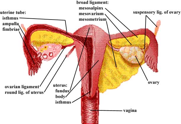
- Describe the broad ligament and differentiate its parts.
- Identify the ovary and discuss the functional significance of its ligaments.
- Demonstrate the uterine tube and its subdivisions.
- Identify the uterus and its subdivisions and demonstrate the continuity of its lumen with that of the uterine tubes and the vagina.
- Differentiate between the internal and external os of the cervix.
- Identify the vagina, and note the angle formed at its junction with the uterus.
- Trace the entire course of the ductus deferens and identify its ampulla; note its relationship to the ureter.
- Identify the seminal vesicle and demonstrate the formation and course of the ejaculatory duct.
- Identify the prostate gland and describe the special features of the prostatic urethral wall.
- Identify the testis, its coverings, and tubules, and account for the difference in location between gonads in the two sexes.
- Demonstrate the epididymis and its subdivisions.
Learning Objectives and Explanations:
1. Trace the continuity of the abdominal peritoneum with that of the pelvic cavity, and identify the peritoneal pouches of the pelvic floor in both sexes. (W&B 533-534, N 360, 361, 362, 363, 371, TG 6-07A, 6-07B, 6-08A, 6-08B, 6-11, 6-13)2. Identify the superficial features of the external genitalia. (W&B 519-522, N 351, 377, 382, 387, 390, 398, TG 6-02, 6-25A, 6-25B, 6-31)The peritoneum continues from the abdominal cavity into the pelvic cavity, but does not entirely invest the pelvic viscera. In the female, the peritoneum:
- extends from the anterior abdominal wall to the superior surface of the bladder, not drooping low enough to catch the anterior surface
- sweeps over the fundus and covers part of the posterior surface of the bladder
- jumps from the posterior surface of the bladder to the anterior (vesicle) surface of the uterus
- sweeps superiorly, to the fundus of the uterus, contacting the uterine tubes
- the space created by the peritoneum sweeping down the back of the bladder, over to the uterus, and up the front of the uterus is the vesicouterine pouch
- continues around the fundus of the uterus, and over the uterine tubes, to the posterosuperior (intestinal) surface of the uterus
- the "doubling" of the peritoneal layers as they hang on either side of the uterine tubes creates the broad ligaments
- jumps from the uterus to cover the anterior portion of the rectum, starting about 2/3 of the way down the rectum
- continues up the rectum, investing the sides as well as it reaches the superior 1/3, attaching to the posterior body wall
- the space created by the peritoneum sweeping across the uterus, jumping to the rectum, and beginning to travel up the front of the rectum is the rectouterine pouch
3. Recognize the features of the rectum that differentiate it from the colon. (W&B 485, 535, N 307, 311, 360, 361, 393, 394, 409, 410, 412, TG 6-08A, 6-08B, 5-14, 6-15A, 6-15B, 6-15C, 6-16, 6-19A, 6-19B, 5-28)Sorry to cop out and paste in the tables, but there is little connectivity or functionality that needs to be explained. It is pretty much a "these are the things you should know about" situation.
4. Describe the point at which the anal canal begins. (W&B 536-538, N 393, 394, 398, 399, 402, 406, 407, 408, 410, 411, TG 6-16, 6-17A, 6-17B, 6-19A, 6-19B, 5-28, 6-30, 6-33, 6-34)There are two differences visible from the outside. First, the lower one-third of the rectum has nothing to do with peritoneum, and its superior two-thirds is retroperitoneal to varying degrees. The sigmoid colon, on the other hand, is peritoneal throughout its length. Second, the pattern of the teniae coli changes as the transition from sigmoid colon to rectum occurs. The sigmoid colon, like the rest of the colon, has three longitudinal muscular bands. These coalesce into two bands, anterior and posterior, on the rectum.
On the inside, the rectum has side-to-side "waviness", due to transverse rectal folds, that is not present in the sigmoid colon.
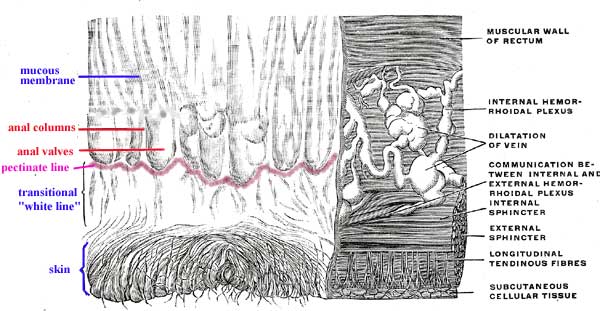
5. Describe the internal features of the anal canal, and determine the point at which its lining changes from cutaneous to mucosal. (W&B 537-538, N 393, 394, 398, 399, 402, 406, 407, 408, 410, 411, TG 6-16, 6-17A, 6-17B, 6-19A, 6-19B, 5-28, 6-30, 6-33, 6-34)The anal canal begins where rectal ampulla narrows, which is at the level of a "U"-shaped sling formed by puborectalis muscle. As a result of this sling, the direction of the gut tube abruptly changes. While the distal rectum runs anteroinferiorly, the anal canal, deviating almost 90 degrees, runs posteroinferiorly.
Images from "Anatomy of the Human Body" by Henry Gray are provided by:
The lining of the anus officially changes from "skin" to mucosal at the pectinate line, though there is a transitional zone, or "white line", between the two areas. The pectinate line runs along the inferior borders of the anal valves, which are mucosal folds connecting the anal columns to one another.
6. Recall the lymph node groups that drain the anal canal. (W&B 535-538, N 266, TG 6-33, 6-34)7. Organize blood and nerve supply to the anal canal. (W&B 535-538, N 393, 394, 398, 399, 402, 406, 407, 408, 410, 411, TG 6-16, 6-17A, 6-17B, 6-19A, 6-19B, 5-28, 6-30)The lymphatic drainage of the anal canal mostly follows the blood vessels supplying the area. There are two primary paths for lymph leaving the area above the pectinate line:
- to the inferior mesenteric lymph nodes - efferent vessels from here go to the superior mesenteric lymph nodes, then to the lumbar nodes/trunk, finally ending in the thoracic duct/cisterna chyli.
- to the internal iliac lymph nodes - efferent vessels from here go to the common iliac lymph nodes, then to the lumbar nodes/trunk, finally ending in the thoracic duct/cisterna chyli.
Lymph from the cutaneous region of the anus, i.e. below the pectinate line, drains to the superficial inguinal lymph nodes. From there it goes to the external iliac and/or deep inguinal lymph nodes, then on to the common iliac lymph nodes, lumbar nodes/trunk, and thoracic duct (cisterna chyli).
8. Recognize the urinary bladder in either its expanded or contracted position, and determine the extent of its peritoneal covering. (W&B 538-539, 541 (fig), 545 (fig), N 360, 361, 362, 366, 402, 403, 406, 407, 408, 410, TG 6-07A, 6-07B, 6-08A, 6-08B, 6-10A, 6-10B, 6-17A, 6-17B, 6-19A, 6-19B, 6-33, 6-34)In addition to dividing the lymphatic drainage, the pectinate line also divides the arterial supply, venous drainage, and innervation of the anal canal. Superior to the pectinate line we have blood coming from the superior rectal artery, blood draining to the portal system via the superior rectal veins, and visceral (no localized pain) innervation via the inferior hypogastric plexus. Inferior to the pectinate line we have blood coming from the inferior rectal arteries, draining into the caval system via the middle and inferior rectal veins (this is an area of anastomosis between the two systems), and somatic (voluntary muscle, cutaneous sensory) innervation via the inferior rectal nerves. (Remember also that there are differences in lymphatic drainage.
9. Identify the internal orifices of the bladder and differentiate the trigone region from the rest of the bladder lining. (W&B 539-540, N 360, 361, 362, 366, 402, 403, 406, 407, 408, 410, TG 6-10A, 6-10B)The superior surface of the bladder is covered by peritoneum as well as the uppermost one or two centimeters of the posterior aspect. The rest of it is extraperitoneal, actually below the lowest extent of the peritoneum.
10. Describe the relationships of the bladder to other pelvic organs in both sexes. (W&B 533-534, N 360, 361, 362, 366, 402, 403, 406, 407, 408, 410, TG 6-07A, 6-07B, 6-08A, 6-08B)There is an "orifice of the ureter" for each ureter, in addition to an internal urethral orifice, for a total of three. These orifices mark the apices of an equilateral triangle, the vesicle trigone, made of smooth mucous membrane. In an undistended bladder, the ureteric orifices lie about three centimeters apart, with the internal urethral orifice between and inferior to them.
11. Describe the normal position and relationships of all organs of the reproductive tracts in both sexes and the role of each in reproductive processes. (W&B 543-553, N 360, 362, 369, 370, 371A, 371B, 378, 382A, 382B, 383, 399, 400, 402, 404A, 404B, 352, 359, 361A, 361B, 362, 363, 365, 384A, 384B, TG 5-34, 6-07A, 6-07B, 6-08A, 6-08B, 6-09A, 6-09B, 6-10A, 6-10B, 6-11, 6-12, 6-14, 6-15, 6-17, 6-23, 6-29, 6-31)The bladder lies in the anterior half of the pelvis, bounded anteriorly and laterally by the pubic symphysis. Posterior to it we have:
- female: vesicouterine septum (pouch), vagina, and uterus (also somewhat superior to the bladder)
- male: rectovesicular septum (pouch), rectum, ductus deferens, and seminal vesicles
Inferior to the bladder we find the pelvic diaphragm (in females) or the prostate gland (in males).
12. Describe the broad ligament and differentiate its parts. (W&B 547-548, N 371, TG 6-07, 6-08, 6-11, 6-12)These diagrams will be useful here, and for the remaining objectives in this session:
Female:
Male:
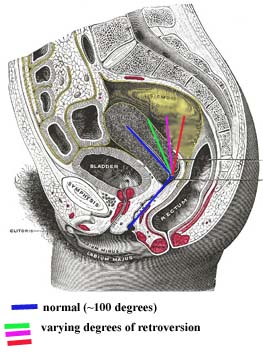
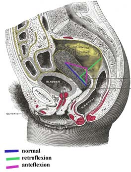
Female: The uterus is more or less horizontal in orientation. It actually lies upon the bladder, so its vesical surface is partially anterior but mostly inferior. Its intestinal surface faces the pelvic cavity. Deviations from this "flopped over the bladder" position can happen. Terms that are "________version" refer to the angle between the long axis of the uterus and the long axis of the vagina. This angle is usually about 100 degrees. Cases where the uterus stands more vertically, and therefore that angle approaches 180 degrees, are called RETROversion. Terms that are "________flexion" refer to the long axis of the body of the uterus as compared to the long axis of the uterine cervix. The long axis of a normal body of the uterus is relatively horizontal, while in the cervix the axis turns partially vertical. With RETROflexion the uterus is actually flopped back, away from the bladder. With ANTEflexion, the uterus is kinked, with a marked angle between the long axis of its body and the long axis of its cervix. (A slight bit of anteflexion is very normal.) Cases where the entire uterus, cervix and all, is moved posteriorly are called retrocession. The vagina is longer here.
Images from "Anatomy of the Human Body" by Henry Gray are provided by:
The uterine (fallopian) tubes attach somewhat laterally to the uterus. They reach toward the ovaries, but are not "officially" attached to them. The fimbriae of the uterine (fallopian) tubes come closest to contacting the ovary. The ovaries themselves are the female gonads. They lie against the pelvic walls.
The female gametes (ova, eggs) mature in the ovaries. In the normal case, one egg is released per month from one ovary or the other. This egg is actually released into the pelvic cavity, but the waving motion of the fimbriae help guide the egg into the uterine tube. The tube is the usual site of fertilization of the egg by sperm, then the newly formed zygote travels to the uterus. It is the job of the uterus to provide an environment for growth of the embryo.
Male: The normal positioning of things in the male is pretty straightforward. The testes are suspended outside the body within the scrotum. A ductus deferens (vas deferens) leads from each testis, through the inguinal canal, around to the posterior part of the bladder. Here they dilate, forming the ampulla of the ductus deferens. Also on the posterior aspect of the bladder lie the seminal vesicles. On each side, the ductus deferens and seminal vesicle join to form the ejaculatory duct, which dumps into the prostatic portion of the urethra. From there, the urethra travels through the penis to its external opening.
The male gametes (sperm) develop within the tissue of the testes. They leave the testes proper as immature gametes. They finish their maturation and are stored in the epidiymis. When needed, they quickly traverse the entire length of the ductus deferens to the ejaculatory duct. At this junction with the seminal vesicles, important materials are added to the semen, the fluid surrounding the sperm. The prostate gland also makes a contribution to the semen. From here the whole mix is sent down the urethra, through the penis, and out of the body.
13. Identify the ovary and its two ligaments and tell the functional significance of both ligaments. (W&B 547-548, N 369, TG 6-07, 6-08, 6-11, 6-12)The broad ligament is a section of peritoneum, like a mesentery, which extends from the pelvic walls to the uterus and uterine (fallopian) tubes. Its three parts are continuous with each other, making it difficult to discern at their junctions. The mesosalpinx is the peritoneum that covers the uterine tube and hangs below it to meet with the mesovarium. The mesovarium is the peritoneum covering the ovary and ovarian ligament, extending like a shelf posteriorly from the mesosalpinx. (If you squint at the diagram in #11, you'll see a ridge of peritoneum running along the top of each ovary.) The mesometrium is the rest of the broad ligament - all of the peritoneum directly connected to the uterus and extending toward the lateral abdominal wall. (Greek, metra = uterus, from meter (mother);
14. Demonstrate the uterine tube and its subdivisions. (W&B 547-548, N 371, TG 6-07, 6-08, 6-11, 6-12)The suspensory ligaments of the ovaries are peritoneal folds covering the ovarian neurovascular supply as the vessels pass over the pelvic brim and into the pelvis to reach the ovary. The suspensory ligament conducts ovarian arteries and veins, nerves, and lymphatics to the ovary. The "ovarian ligament" proper is a round cord which attaches the ovary to the uterus just below the entrance of the uterine tube into the uterus. The ovarian ligament, a remnant of a portion of the gubernaculum, is within the mesovarium.
15. Identify the uterus and its subdivisions and demonstrate the continuity of its lumen with that of the uterine tubes and the vagina. (W&B 548-551, N 371, TG 6-07, 6-08, 6-11, 6-12)The uterine tube extends laterally about 10 cm from the uterus to the ovary. It has three parts:
- isthmus: the constricted part adjacent to the uterus
- ampulla: the widest and longest part, extending laterally to the infundibulum from the isthmus
- infundibulum: the funnel-like terminus, with fringed processes called fimbriae that contact the ovary.
16. Differentiate between the internal and external os of the cervix. (W&B 551-553, N 371, TG 6-11, 6-12)The lumen of the uterus is continuous on both sides with the lumens of the uterine tubes. It is continuous inferiorly with the lumen of the vagina. It is divided into four parts:
17. Identify the vagina, and note the angle formed at its junction with the uterus. (W&B 551-553, TG 6-08, 6-11, 6-12, 6-13)The tapered neck or cervix of the uterus is traversed by the cervical canal. Above, it is continuous with the cavity of the body of the uterus at the internal os. Below, at a depression on the vaginal portion of the cervix, the external os opens into the cavity of the vagina.
18. Trace the entire course of the ductus deferens and identify its ampulla; note its relationship to the ureter. (W&B 545, N 361A, 361B, 363, 384A, 384B, 390, TG 6-10, 6-31A, 6-31B)The vagina is muscular, but not as much as the uterus. The angle between its long axis and the long axis of the uterus is about 100 degrees in normal cases. Other cases can occur, as described in #12 above.
19. Identify the seminal vesicle and demonstrate the formation and course of the ejaculatory duct. (W&B 545-546, N 361A, 361B, 384A, 384B, TG 6-10)The ductus deferens is an unbelievable 45 cm long! It starts in the tail of the epididymis, ascends as part of the spermatic cord, traverses the inguinal canal, emerges from the deep inguinal ring, passes lateral to the inferior epigastric artery, ascends obliquely across the external iliac arteries, goes over to the back side of the bladder and descends on the fundus medial to the ureter and seminal vesicles. The ductus deferens joins the duct of the seminal vesicle at the prostate to form the ejaculatory duct. The most distal portion of the ductus is the ampulla, which is tortuous and dilated. ("Ductus deferens" literally means "deferent vessel", which means "vessel that shows respect and esteem due a superior or an elder".)
20. Identify the prostate gland and describe the special features of the prostatic urethral wall. (W&B 546-547, N 361, 384A, 384B, TG 6-10)The seminal vesicles lie lateral to the ampulla of the ductus deferens on the posterior side of the bladder. Their ducts join the ductus deferens to form the ejaculatory ducts. They are similar in structure to the ampulla of the ductus.
21. Identify the testis, its coverings, and tubules, and account for the difference in location between gonads in the two sexes. (W&B 431-433, 544-545, N 387, 388, 390, TG 5-10, 6-31, 6-32)The prostate gland sits under the bladder, with the ampulla of the rectum posterior to it. (The rectovesical septum is between the two.) The base of the gland, which is the TOP, is continuous with the bladder wall. The ejaculatory ducts enter the prostate posterosuperiorly and run to the prostatic urethra.
The prostatic urethra is three to four centimeters long. The urethral crest is a ridge within it, and the prostatic sinuses are valleys on either side of the crest. The little ducts of the prostate gland empty into these sinuses.
The seminal colliculus is a rounded eminence in the middle of the prostatic urethra. A small slit in this structure leads to a very small blind pouch in the prostate. This pouch is the prostatic utricle (or "vagina masculina"), the male homologue of the uterus and vagina, i.e. the remnant of the Mullerian duct system. Near the mouth of the prostatic utricle, the two ejaculatory ducts empty into the prostatic urethra. (Latin, utriculus = little leather bag)
22. Demonstrate the epididymis and its subdivisions. (W&B 544, N 390, TG 6-32)The testes are covered by a tough, fibrous coat called the tunica albuginea testis. Superficial to that they are invested by two layers of tunica vaginalis testis, much like the pleura or peritoneum. (This structure is actually derived from the processus vaginalis of the peritoneum.) The visceral layer of the tunica vaginalis testis almost completely covers each, only being absent where the testis attaches to the epididymis. The parietal layer of the tunica vaginalis testis is more extensive yet, extending for a short distance along the spermatic cord. (Latin, tunica albuginea = white membrane, covering)
The gonads have a similar path of development in both sexes, up to a point. That point is when the ovaries try to start descending and find that the uterus is in the way. So, they stay where they are.
The epidiymis is a coil of tubes that lies on the posterior aspect of the testis. (The total length of each tube, if uncoiled, is 15-20 feet.) It is divided into three parts: (Greek, epi = upon, didymos = testicle, twin)
- head of the epididymis: the largest mass of coils, located on the superoposterior part of the testis
- body of the epididymis: inferior to the head, the coiling here is not as pronounced
- tail of the epididymis: the smallest, most inferior portion, which connects to the ductus deferens
Cultural enrichment: Check out these sections from the 1918 version of Gray's Anatomy of the Human Body! Some of the terms are (of course) out-of-date, but the illustrations are timeless.
Questions and Answers:
23. Note the difference between male and female in the subpubic angle, the angle formed by the subpubic arch. What are other sex differences in the pelvic skeleton? (W&B 571-573, N 354, TG 6-05A, 6-05B, 6-05CD, 6-05EF)24. Define the rectum. (N 393, 394, TG 6-08A, 6-08B, 6-15A, 6-15B, 6-15C, 6-16)
Structure/Section Female Male pelvic inlet oval and rounded heart-shaped pelvic outlet large small pubic arch and subpubic angle wide narrow iliac wings flared less flared 25. Define and note the flexure between rectum and anal canal. What muscle assists in maintaining this flexure? (N 393, TG 6-08A, 6-08B, 6-15A, 6-15B, 6-15C, 6-16)The rectum begins where the peritoneal investment of the sigmoid ends, at approximately the level of S3. It curves into the pelvic diaphragm, dilating just above the pelvic diaphragm as the rectal ampulla, and ends four centimeters below and in front of the coccyx. It continues as the anal canal after piercing the pelvic diaphragm. The rectum is about twelve centimeters long altogether. Its anterior surface contacts the vagina in females or the prostate gland in males.
26. In the mucosa, define the anal columns and the pectinate line. What is their significance? (N 393, TG 6-16)The anterior flexure at the anorectal junction is held by the sling of the puborectalis portion of the levator ani muscle, which passes posteriorly around the anorectal junction.
27. On the sagittally-sectioned female specimen, trace the peritoneum from the ventral abdominal wall; examining the vesicouterine pouch and its manner of reflection from the bladder to the uterus. Onto what part of uterus does it reflect? (N 378, TG 6-08A)See #8 above.
28. Trace the peritoneum across the uterus and define the rectouterine pouch. Note peritoneum on the posterior wall of the vagina. From what point does the peritoneum reflect to the rectum? What is the significance of this? (N 360, TG 6-08A)The peritoneum on the superior surface of the bladder reflects onto the uterus at the isthmus, just superior to the cervix.
29. Within the broad ligament, locate the ovarian ligament and the round ligament of the uterus. Consider development and continuities of these structures. (N 367, 420, TG 6-11, 6-12)The peritoneum of the rectouterine pouch lies in contact with the posterior fornix of the vagina. This allows incisions, punctures, or lacerations of the posterior fornix of the vagina to open the peritoneal cavity. (This is often how eggs are harvested these days.)
30. Locate and define the peritoneal fold called the suspensory ligament of the ovary. What does it contain? (N 374, 400, TG 6-11A, 6-11B, 6-12)The proper ovarian ligament and the round ligament of the uterus are both remnants of the gubernaculum. They are continuous with one another where they contact the lateral surface of the uterus inferior to the uterine tube.
31. Strip the peritoneum from the suspensory ligament of the ovary on one side and trace the ovarian artery and vein. What are their sources? (N 400, TG 5-34)The suspensory ligament of the ovary contains ovarian vessels, autonomic nerves, lymphatics, and extraperitoneal connective tissue.
32. What is the complete area of distribution of the ovarian artery? (N 400, TG 6-11B, 6-12)The ovarian artery branches from aorta. The right ovarian vein drains to inferior vena cava. The left ovarian vein drains to left renal vein. (This is analogous to testicular vessels in males.)
33. Locate a ureter. Note its relation to uterine artery. Trace it to the bladder and posteriorly to the brim of the pelvis, noting course, relation to peritoneum, and blood supply. (N 400, TG 6-11B, 6-17)The ovarian artery supplies the ovary, mesovarium, and infundibulum of the uterine tube.
34. Trace the round ligament from the uterus to the deep inguinal ring. Where does it attach? (N 369, TG 6-07A, 6-11B, 6-12)The ureter passes over the pelvic brim just medial to the ovarian vessels, usually at the bifurcation of the common iliac artery. The ureter then descends and passes anteriorly within the pelvis. It is crossed superiorly by the uterine artery ("bridge over water") before it turns medially to enter the posterior wall of the bladder.
35. What structures support the uterus? (N 369, TG 6-13E)The round ligament of the uterus attaches to the lateral surface of uterus, below and anterior to the intramural portion of the uterine tube. It helps to hold the fundus of the uterus forward (anteverted).
36. Examine the vagina and the structure of its wall. Consider differences between the vagina and the vestibule of the vagina. (N 370, TG 6-08, 6-11, 6-12, 6-13)The round ligament of the uterus helps to hold the uterus in an anteverted position superior to the bladder. The cardinal ligaments and the endopelvic fascia around the uterine vessels helps to fix the cervix in place, as do the rectouterine ligaments. Even the broad ligament lends a slight amount of support to the uterus. (Anterversion of the uterus seems to be key, since retroversion is associated with prolapse of the uterus into the vagina. See #12 above.)
37. Examine the intravaginal cervix, the ostium of the uterus, and fornices of the vagina. Note relations to urethra, bladder, and rectum. What is the significance? (N 360, 370, 371, TG 6-08, 6-11, 6-12, 6-13)The vestibule of the vagina is the cleft between the paired labia minora. The vagina proper extends from the hymen or hymeneal caruncles at the vaginal orifice superiorly to the cervix of the uterus. Its wall is relatively muscular and covered with mucosa.
38. Explore the female urethra, noting length, sphincter muscle, and relation to vagina. Note specifically the relation of the orifice to the anterior vaginal wall. What is the significance? (W&B 549, N 383, 379, TG 6-08, 6-10)See descriptions of peritoneum, urinary apparatus, and vagina above.
39. Define the ampulla of the ductus deferens. Is it covered by peritoneum? (N 361A, 361B, 384A, 384B, TG 6-14)The urethra is about four centimeters long. It is homologous to male prostatic/membranous urethra. Its orifice is within the vestibule of the vagina, immediately in front of the vaginal orifice.
40. What is the rectovesical pouch? (N 361A, 361B, TG 6-08)The ductus deferens is covered by peritoneum, but its ampulla is not. The upper end of the seminal vesicle may contact peritoneum, otherwise it is inferior to the peritoneum lining the rectovesical pouch.
41. Where does the transition of the epididymis to the ductus deferens occur? (N 390, TG 6-32)The rectovesical pouch is the reflection of peritoneum between the rectum and the bladder. This of course only occurs in males.
It occurs at the tail of the epididymis on the posteroinferior aspect of the testis.