|
|
|
||||||||||||
Dissector Answers - Pelvic Neurovasculature |
|||||||||||||
Learning Objectives:
Upon completion of this session, the student will be able to:
- Describe the formation of the two sciatic foramina. List the muscles, nerves, and vessels which pass through each.
- Demonstrate the origins of the piriformis and obturator internus muscles and describe two specializations of the obturator fascia.
- Identify the pelvic diaphragm and differentiate its components.
- Trace the branching pattern of the internal iliac vessels in each sex, identifying branches by their relationships to pelvic organs or wall structures.
- Demonstrate the formation of the sacral plexus, its relationship to the piriformis muscle and gluteal vessels, and its pelvic splanchnic nerves.
- Identify and describe the inferior hypogastric (pelvic) plexus and its connections to the superior hypogastric plexus via the hypogastric nerves.
- Identify and describe the sacral sympathetic trunks and the sacral sympathetic nerves.
- Trace the sympathetic and parasympathetic nerve supply to any pelvic organ, listing the location of the preganglionic cell body, the course of preganglionic fibers, the location of the postganglionic cell body, and the course of postganglionic fibers.
Learning Objectives and Explanations:
1. Describe the formation of the two sciatic foramina. List the muscles, nerves, and vessels which pass through each. (W&B 570-571, N 352, 503, TG 3-28, 6-06)2. Demonstrate the origins of the piriformis and obturator internus muscles and describe two specializations of the fascia of the latter. (W&B 564-565, N 367A, 367B, 369, 503, TG 3-28, 6-21A, 6-21B, 6-22)This is something that only makes sense in three dimensions. You must look at a pelvis from your bone box or in the lab, and figure out where these ligaments are going. Luckily, they are named in a very logical way. These anterior and posterior views might help.
Images from "Anatomy of the Human Body" by Henry Gray are provided by:
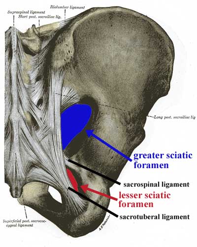
The sacrotuberal ligament connects the sacrum to the the ischial tuberosity. With the pelvis in the correct position, it runs mostly inferolaterally from the sacrum to the tuberosity, and only slightly anteriorly. The sacrospinal (sacrospinous) ligament connects the sacrum to the ischial spine. With the pelvis in correct anatomical position, it runs anterolaterally from the sacrum to the ischial spine, but does not deviate much in the superior-inferior axis.
These ligaments, along with the greater sciatic notch and the lesser sciatic notch, make up the greater sciatic foramen and the lesser sciatic foramen, respectively.
greater sciatic foramen: bounded anteriorly and superiorly by the posterior border of the hip bone (greater sciatic notch), posteriorly by the sacrotuberal ligament, and inferiorly by the sacrospinal ligament. The piriformis muscle passes through this opening, as do these nerves and vessels:
- superior to piriformis muscle: superior gluteal vessels and nerve
- inferior to piriformis muscle: inferior gluteal vessels and nerve, the sciatic nerve, the posterior femoral cutaneous nerve, and the nerve to the quadratus femoris muscle - also, the internal pudendal vessels and nerve and the nerve to the obturator internus muscle leave the pelvis via this opening, but enter the perineum through the lesser sciatic foramen (see below)
lesser sciatic foramen: bounded anteriorly by the ischial tuberosity, superiorly by the ischial spine and sacrospinal ligament, and posteriorly by the sacrotuberal ligament. It transmits the tendon of the obturator internus muscle - also, the nerve to the obturator internus muscle and the internal pudendal vessels and nerve, which left the pelvis via the greater sciatic foramen, re-enter the pelvis (in the case of the nerve to the obturator internus muscle) or the perineum (in the case of the internal pudendal vessels and nerve) via the lesser sciatic foramen
3. Identify the pelvic diaphragm and differentiate its components. (W&B 565-566, N 367A, 367B, 368, 369, 370, TG 6-23, 6-21A, 6-21B, 6-22, 6-23)The piriformis muscle takes origin from the anterior surfaces of S2 to S4, both between and lateral to the sacral foramina. It exits the pelvis via the greater sciatic foramen, inserting on the greater trochanter of the femur in order to rotate the thigh laterally. (Latin, piriformis = pear-shaped)
The obturator internus muscle takes origin from the whole bony rim of the obturator foramen, the inner surface of the obturator membrane, and a large area of the inner surface of the ischium. It leaves the pelvis (or, more correctly, its tendon does so) via the lesser sciatic foramen to insert onto the greater trochanter of the femur in order to rotate the thigh laterally. Its fascia has two specializations. First, there is a strong band that stretches between the spine of the ischium and the superior pubic ramus. This is the arcus tendineus levator ani, which gives origin to the levator ani muscles. The other specialization is the obturator membrane, which nearly covers the entire obturator foramen, only leaving space for the obturator nerves and vessels to exit.
4. Trace the branching pattern of the internal iliac vessels in each sex, identifying branches by their relationships to pelvic organs or wall structures. (W&B 553-559, N 398, 400, 401, 402, 403A, 403B, 404A, 404B, 405, TG 6-17A, 6-17B, 6-29A, 6-29B)The pelvic diaphragm is shaped like a bowl, with bony attachments at the pubic symphysis and the coccyx. Between those bones, it is attached to a thickening of the obturator internus muscle fascia called the arcus tendineus levator ani. It is made of two muscles, one of which is divided into 3 (or 4) parts.
Muscle Origin Insertion Action coccygeus ischial spine side of the coccyx and lower sacrum elevates the pelvic floor levator ani posterior surface of the body of the pubis, arcus tendineus levator ani, ischial spine anococcygeal raphe and coccyx elevates the pelvic floor iliococcygeus (part of levator ani) arcus tendineus levator ani and the ischial spine anococcygeal raphe and the coccyx elevates the pelvic floor pubococcygeus (part of levator ani) posterior aspect of the superior pubic ramis coccyx elevates the pelvic floor puborectalis (part of levator ani) posterior aspect of the body of the pubis unites with the puborectalis m. of other side posterior to the rectum draws the distal rectum forward and superiorly pubovaginalis (sometimes listed, part of levator ani) posterior aspect of the body of the pubis fascia of the vagina and perineal body draws the vagina forward and superiorly Furthermore, the diaphragm includes the inferior fascia and the superior fascia. The inferior fascia is an extension of the obturator internus fascia and is continuous with the fascia of the external sphincter ani muscle. The superior fascia is an extension of the transversalis fascia. The two layers are continuous in the urogenital hiatus.
5. Demonstrate the formation of the sacral plexus, its relationship to the piriformis muscle and gluteal vessels, and its pelvic splanchnic nerves. (W&B 559-561, N 402, 409, 410, 412, 415, 416, 417, 497, 499A, 499B, TG 3-25A, 3-25B, 6-17, 6-18, 6-19)The abdominal aorta splits into two common iliac arteries at the level of the L4 vertebra. The common iliac arteries each give off external and internal branches at about the level of the disk between L5 and S1. The external iliac arteries continue outside of the pelvis to supply the lower extremities. The internal iliac arteries enter the pelvic cavity, and generally divide into anterior and posterior divisions to supply pelvic viscera, the buttocks, some of the medial thigh, and the perineum.
The internal iliac artery system is one of the most variable in the body. Therefore, don't try to memorize the exact order of the branches. Just know which division they usually come from and any spatial relationships that are important.
Each posterior division only gives off branches to muscle and body wall. These include:
- superior gluteal artery: supply to gluteus maximus muscle, gluteus medius muscle, gluteus minimus muscle, and hip joint
- iliolumbar artery: supply to iliacus muscle, psoas major muscle, and quadratus lumborum muscle via iliac and lumbar branches
- lateral sacral arteries (usually two per side): supply to sacrum, sacral nerve rootlets, meninges, and adjacent muscles
Each anterior division gives off branches to muscle and body wall, as well as branches to pelvic viscera. Branches to muscle and body wall include:
- inferior gluteal artery: supplies gluteus maximus muscle and the hip joint
- obturator artery: supply to the medial thigh and hip via pubic, acetabular, anterior, and posterior branches
- internal pudendal artery: the primary blood supply to the perineum. It supplies the anus, muscles of the superficial and deep perineal spaces, clitoris or penis, and the posterior aspect of the labium majus or scrotum via numerous branches. (Latin, pudere = to be ashamed)
Branches to pelvic viscera from the anterior division include:
- middle rectal artery: supply to the middle portion of the rectum
- umbilical artery: supply to the superior part of the bladder by giving off the superior vesical arteries, and, in males, to the ductus deferens via the artery of the ductus deferens. (Distal to those branches, the umbilical artery is not patent, and becomes the medial umbilical ligament.)
- inferior vesical artery: supply to lower part of the bladder and to the vagina or prostate. It anastomoses with, and sometimes is a branch of, the middle rectal artery.
- uterine artery (in females): supply to uterus, uterine tube, and upper vagina. The latter two are via tubal and vaginal branches.
- vaginal artery (in females): supply to a major portion of the vagina. It anastomoses with, and sometimes is a branch of, the uterine artery.
For more than you ever wanted to know about anatomical variation, the University of Iowa has a great site, an "Illustrated Encyclopedia of Human Anatomic Variation". Here is a quick and dirty link to the section on internal iliac arteries.
The internal ilac vein on each side receives tributaries that roughly correspond to the branches of the internal iliac artery. These include the superior and inferior gluteal veins, the obturator vein, the internal pudenadal vein, the lateral sacral vein, the middle rectal vein, the vesical vein, prostatic vein (in males), and/or uterine and vaginal veins (in females). The iliolumbar vein drains directly into the common iliac vein on each side.
6. Identify and describe the inferior hypogastric (pelvic) plexus and its connections to the superior hypogastric plexus via the hypogastric nerves. (W&B 562-564, N 409, 410, 412, TG 8-18, 8-19, 8-20, 8-21)Images from "Anatomy of the Human Body" by Henry Gray are provided by:
The sacral plexus proper includes contributions from L4 through part of S4. (Also shown here is the coccygeal plexus, which includes the rest of S4, S5, and the coccygeal nerve.) All of the contributing nerves, except S4, divide into anterior (shown yellow above) and posterior (shown yellow with black shading above) branches.
sciatic nerve: actually two separate nerves that are usually fused together (though in some cases they are not) - the tibial nerve, from anterior branches of L4 through S3, and the common fibular (peroneal) nerve, from the posterior branches of L4 through S2. The sciatic nerve runs under the piriformis muscle, emerging at its inferior border. It travels through the greater sciatic foramen, and on into the thigh. The sciatic nerve provides motor innervation to the hamstrings (posterior thigh muscles) and all of the muscles in the leg and the foot.
superior gluteal nerve: from posterior branches of L4 through S1. Travels through the greater sciatic foramen superior to the piriformis muscle, and moves laterally between the gluteus medius muscle and the gluteus minimus muscle. Provides motor innervation to gluteus medius muscle, gluteus minimus muscle, and tensor fascia lata muscle.
inferior gluteal nerve: from the posterior branches of L5 through S2. Travels through the greater sciatic foramen inferior to the piriformis muscle, and travels for a short distance deep to the gluteus maximus muscle but superficial to the sciatic nerve. It provides the sole motor innervation of the gluteus maximus muscle.
nerve to quadratus femoris muscle: from the anterior branches of L4 through S1. Travels through the greater sciatic foramen inferior to the piriformis muscle. Provides motor innervation to quadratus femoris and inferior gemellus muscles.
nerve to obturator internus muscle: from the anterior branches of L5 through S2. Travels through the greater sciatic foramen inferior to the piriformis muscle, but then loops back around to re-enter the pelvis through the lesser sciatic foramen. Provides motor innervation to the obturator internus and superior gemellus muscles.
posterior femoral cutaneous nerve: from the anterior branches of S2 and S3 and the posterior branches of S1 and S2. Travels through the greater sciatic foramen inferior to the piriformis muscle, and lies alongside the sciatic nerve. It provides sensory innervation to the skin of the back of the thigh in addition to the skin of the lower and lateral buttocks (through gluteal branches) and the perineum (via perineal branches).
pudendal nerve: from the anterior branches of S2 through S4. Travels through the greater sciatic foramen inferior to the piriformis muscle, but then loops back around to enter the perineum through the lesser sciatic foramen, entering the pudendal canal. Provides motor innervation to the muscles of the perineum, and is the primary sensory innervation to the genitalia. (Latin, pudere = to be ashamed)
perforating cutaneous nerve: from the posterior branches of S2 and S3. Pierces the sacrotuberal ligament to provide sensory innervation to the skin of the medial part of the fold of the buttock.
nerve to piriformis muscle: from posterior branches of S1 and S2. Provides motor innervation to the piriformis muscle.
nerves to coccygeus and levator ani muscles: from anterior branches of S3 and S4. Provide motor innervation to coccygeus and levator ani muscles.
pelvic splanchnic nerves: from the ventral primary rami of S2 through S4. Travel to the inferior hypogastric plexus (see below).
perineal branch of the fourth sacral nerve: from the anterior branch of S4. Descends through the coccygeus muscle and runs anteriorly to the external sphincter ani muscle. Provides motor innervation to this muscle and sensory innervation to the overlying skin.
7. Identify and describe the sacral sympathetic trunks and the sacral splanchnic nerves. (W&B 562-564, N 415, 416, 417, TG 8-18, 8-20)The inferior hypogastric plexus is a major meshwork of nerves that are located on either side of the rectum, cervix, and lateral vagina in the female, or on either side of the rectum, prostate, and seminal vessicles in the male. It receives the following:
hypogastric nerves: from the superior hypogastric plexus. These are the primary ways in which sympathetic neurons reach the hypogastric plexus, and therefore the pelvic viscera.
sacral splanchnic nerves: from the second and/or third ganglia of the sacral sympathetic trunk. These are the secondary ways in which sympathetic neurons reach the hypogastric plexus, and therefore the pelvic viscera.
pelvic splanchnic nerves: from the ventral primary rami of S2 through S4. These are the ways in which parasympathetic neurons reach the hypogastric plexus, and therefore the pelvic viscera and distal colon. (Remember that the parasympathetic part of the autonomic nervous system is the "craniosacral" part. Parasympathetic innervation to most of the gut comes from the "cranio-" half of that, i.e., the vagus nerve. The rest, to colon distal to the splenic flexure and to pelvic viscera, is from the "-sacral" half, via the pelvic splanchnic nerves. See also #7 below.)
Note: So far we have seen thoracic, lumbar, sacral, and pelvic splanchnic nerves. Remember that "splanchnic" really only means that they are going to viscera. It so happens that the first three emerge from sympathetic chain ganglia and carry sympathetic fibers, while the fourth has nothing to do with the sympathetic nervous system. If it helps, i.e. does not confuse you more, just remember that the splanchnic nerves come from the chain ganglia and carry sympathetic fibers, with the exception of the pelvic splanchnic nerves.
8. Trace the sympathetic and parasympathetic nerve supply to any pelvic organ, listing the location of the preganglionic cell body, the course of preganglionic fibers, the location of the postganglionic cell body, and the course of postganglionic fibers. (TG 8-18, 8-19, 8-20, 8-21)The sacral sympathetic trunk is slender, and often has 4 or fewer visible ganglia. The trunks lie on the anterior surface of the sacrum and the origin of piriformis, medial to the anterior sacral foramina, which have the large sacral ventral primary rami emerging laterally. The trunk has gray rami branching laterally to reach these sacral VPR's, and you may see very slender, hair-like sacral splanchnic nerves passing anteriorly onto the sides of the rectum to join the inferior hypogastric plexus.
Sympathetic: The preganglionic cell body is located in the lateral horn of the spinal cord in the thoracolumbar region. Axon travels out of the cord via the spinal nerve to the sympathetic chain ganglion. In the lumbar region, some of the nerves will synapse there and either travel down the chain to the pelvis or out via the lumbar splanchnic nerves. In the former case, they leave the chain in the pelvis, via a sacral splanchnic nerve, to reach the hypogastric plexus. In the latter case, they may reach the hypogastric plexus (the long way) via the superior hypogastric plexus and hypogastric nerves.
Parasympathetic: The preganglionic cell body is located in the lateral horn of the spinal cord in the sacral region. Axon travels out of the spinal cord via S2, S3, or S4 spinal nerves. These give off pelvic splanchnic nerves, which reach the hypogastric plexus. From here the neurons travel to their target, usually synapsing with a postganglionic neuron within the tissue of the target organ.
Cultural enrichment: Check out these sections from the 1918 version of Gray's Anatomy of the Human Body! Some of the terms are (of course) out-of-date, but the illustrations are timeless.
Questions and Answers:
9. Locate the anterior division of the internal iliac artery and note how it terminates by dividing into the inferior gluteal and the internal pudendal arteries. These exit the pelvis below the lower border of the piriformis muscle. What are other relations? (N 402, 403A, 403B, 502, TG 3-29, 6-17A, 6-17B)The internal pudendal and inferior gluteal (the larger of the two) arteries are terminals of the anterior division of the internal iliac artery. They arise from a common trunk either within or outside the pelvis. The internal pudendal artery exits the greater sciatic foramen between the piriformis and coccygeus muscles, crosses the iliac spine to pass through the lesser sciatic foramen, and enters the pudendal canal. The inferior gluteal artery passes between the second and third sacral nerves to leave the pelvis below the piriformis muscle.10. Do you have an "aberrant obturator artery", which arises from the inferior epigastric artery and accompanies the obturator nerve?An aberrant obturator artery takes its origin from the inferior epigastric or, rarely, from the external iliac itself. It would descend along the brim of the pelvis to the obturator foramen.11. Locate the sympathetic trunk entering the pelvis along the medial border of the pelvic sacral foramina. Note number of ganglia, gray rami communcantes, and sacral splanchnic nerves. (N 410, 412, TG 8-18, 8-20)Both sympathetic trunks descend on the anterior surface of the sacrum in the extraperitoneal connective tissue. There are usually four ganglia in the sacral region, one opposite the upper three sacral segments and one beneath the fourth and fifth segments of the sacrum. The two trunks typically unite over the coccyx to form the "ganglion impar". Sacral splanchnic nerves are slender fibers leaving the anterior surface of the sacral sympathetic trunk ganglia to enter the inferior hypogastric plexus on the sides of the rectum. Gray rami communicantes also leave the lateral side of the sacral sympathetic trunk to reach the sacral ventral primary rami as they emerge from the anterior sacral foramina.12. How many pelvic splanchnic nerves are there? (N 410, 412, TG 8-19, 8-21)The pelvic splanchnic nerves represent the sacral portion of the craniosacral outflow or parasympathetic portion of the autonomic nervous system. The pelvic splanchnic nerves spring from the ventral rami of the second, third, and fourth sacral nerves. The contribution from the third sacral nerve is usually the largest. From three to ten strands of nerves pass forward and become incorporated into the inferior hypogastric plexus.13. What is the puborectalis muscle? What is its significance? (N 367A, 367B, 368, 369, 370, TG 6-21A, 6-21B, 6-22, 6-23A, 6-23B)The puborectalis muscle is the most medial portion of the levator ani muscle. It passes around the terminal rectum to form the puborectal sling, which kinks the anorectal junction forward to assist in maintaining fecal continence. This muscle marks the transition from rectum to anus.14. Define the urogenital hiatus. What does it transmit? (N 367A, 367B, 368, 369, 370, TG 6-21A, 6-21B, 6-22, 6-23A, 6-23B)The passage (transmission) of the urethra/vagina and anus through the pelvis requires a separation of the two halves of the pelvic diaphram in front of the rectum. This is the urogenital hiatus.