|
|
|
||||||||||||
Dissector Answers - Anterior Leg & Foot |
|||||||||||||
Learning Objectives:
Upon completion of this session, the student will be able to:
- Identify the muscles of the anterior and lateral compartments of the leg and give their functional significance in locomotion.
- Identify the vascular supply of the anterior and lateral compartments of the leg.
- Identify the nerves of the anterior and lateral compartments of the leg, the muscles and cutaneous regions supplied by them, so that given a functional and/or cutaneous loss one can predict the nerve and the probable level of injury.
- Describe the bony structure of the foot, including its arches, subtalar and transverse tarsal joints, and the bones and ligaments contributing to its strength and flexibility.
- Describe the arrangement, specializations, and compartments of the foot.
- Identify the muscles of the foot and give their functional significance in locomotion.
- Identify the vascular supply of the foot and give the regions supplied by each.
- Identify the nerves of the foot, and the muscles and cutaneous regions supplied by them, so that given a functional and/or cutaneous loss one can predict the nerve and the probable level of injury.
Learning Objectives and Explanations:
1. Identify the muscles of the anterior and lateral compartments of the leg and give their functional significance in locomotion. (W&B 610-616, TG3-36, TG3-37)2. Identify the vascular supply of the anterior and lateral compartments of the leg. (W&B 618-620, TG3-38, TG3-39A, TG3-39B)The good news about this lab is that muscles are named for what they do, who they do it to, and what they look like. Extensors extend the digits or hallux (the big toe) and are on the front of the leg and foot. Flexors are on the back of the leg and sole of the foot. (Remember, we're talking about the toes being flexed and extended here, not the ankle.) Adduction means toward midline, which is defined as second toe in the foot. Abduction means motion away from midline. Inversion means that you direct the sole of your foot toward midline. Eversion means that you direct the sole of your foot laterally. Plantar flexion means pointing your toe (true flexion at the ankle). Dorsiflexion means bringing the top of the foot up toward the anterior surface of the leg (effectively extension at the ankle). "________ longus" means the muscle is long and you should expect a "_______ brevis", a short muscle that does the same thing. Hallucis means big toe, digitorum refers to the other four toes, and digiti minimi, though not named by Austin Powers, refers to the little toe.
The muscles of the leg are divided into groups, just like the leg is divided into compartments, by the intermuscular septa described above. These are the anterior, lateral, superficial posterior, and deep posterior groups (compartments).
The four muscles of the anterior compartment:
Muscle Origin Insertion Action Innervation tibialis anterior lateral tibial condyle and upper lateral surface of tibia medial surface of medial cuneiform and 1st metatarsal dorsiflexes and inverts foot deep fibular (peroneal) nerve extensor hallucis longus middle half of anterior surface of fibula & interosseous membrane base of distal phalanx of hallux extends metatarsophalangeal & interphalangeal joints of hallux deep fibular (peroneal) nerve extensor digitorum longus lateral condyle of tibia, anterior surface of fibula, lateral portion of interosseous membrane dorsum of lateral 4 toes via extensor expansions (central slip inserts on base of middle phalanx, lateral slips on base of distal phalanx) extends metatarsophalangeal, proximal interphalangeal and distal interphalangeal joints of lateral 4 toes deep fibular (peroneal) nerve fibularis (peroneus) tertius distal part of anterior surface of fibula dorsum of shaft of 5th metatarsal bone everts foot deep fibular (peroneal) nerve Blood supply to the anterior compartment is via the anterior tibial artery.
(The anterior compartment can also be remembered, in order, as "Tom, Harry, Dick AND Fred". That is, medial to lateral you have: Tibialis anterior muscle, extensor Hallucis longus muscle, extensor Digitorum longus muscle, anterior tibial Artery, deep fibular Nerve, and Fibularis tertius muscle.)
The two muscles of the lateral compartment:
Muscle Origin Insertion Action Innervation fibularis (peroneus) longus upper two/thirds of lateral surface of fibula after crossing plantar surface of foot deep to intrinsic muscles, it inserts on medial cuneiform and base of 1st metatarsal plantarflexes and everts the foot superficial fibular (peroneal) nerve fibularis (peroneus) brevis lower one third of lateral surface of fibula tuberosity of base of 5th metatarsal plantarflexes and everts the foot superficial fibular (peroneal) nerve Blood supply to the lateral compartment is via the fibular artery.
(The fibularis brevis muscle is deep to the fibularis longus muscle. Also note that, though the blood supply is via the fibular artery, the fibular artery does not live in the lateral compartment.)
Summary of actions:
Muscles of the anterior compartment of the leg dorsiflex and invert the foot at the ankle. The tibialis anterior muscle is powerful in this action; with the foot on the ground, it draws the tibia forward (as in walking). The extensor hallucis longus muscle dorsiflexes the great toe, while the extensor digitorum longus muscle dorsiflexes toes two through five.
Muscles of the lateral compartment evert and abduct the foot, and also assist in its plantar flexion.3. Identify the nerves of the anterior and lateral compartments of the leg, the muscles and cutaneous regions supplied by them, so that given a functional and/or cutaneous loss one can predict the nerve and the probable level of injury. (W&B 581, 621-622, TG3-39A, TG3-39B, TG3-39A, TG3-68)The popliteal artery is the continuation of the femoral artery in the popliteal fossa. It descends across the popliteus muscle and at its lateral border, divides into the anterior and the posterior tibial arteries. The anterior tibial artery punches forward above the interosseous membrane and into the anterior compartment of the leg. The artery descends on top of the membrane, joined by the deep fibular nerve, and crosses the ankle midway between the lateral and medial malleous. It enters the dorsum of the foot as the dorsalis pedis.
The dorsalis pedis has many branches, and eventually becomes the deep plantar artery. This artery dives to the sole of the foot (between the 2 heads of the 1st dorsal interosseous muscle, between the 1st and 2nd toes). It unites with the lateral plantar artery to form the plantar arterial arch.
The posterior tibial artery gives off the fibular (peroneal) artery and then descends in the deep posterior compartment of the leg, accompanied by the tibial nerve. It passes behind the medial malleolus of the ankle and into the sole of the foot where it divides into the medial and lateral plantar arteries. (Similarly, the tibial nerve divides into medial and lateral plantar branches here.) The medial plantar artery runs in the groove between the medial and central compartments.
4. Describe the bony structure of the foot, including its arches, subtalar and transverse tarsal joints, and the bones and ligaments contributing to its strength and flexibility. (W&B 644-652, TG3-40A, TG3-40B, TG3-41A, TG3-41B, TG3-61B)With respect to specific muscles, the nerves are listed above in #1. In general, they are divided thus:
- anterior compartment - deep fibular (peroneal) nerve
- lateral compartment - superficial fibular (peroneal) nerve
- posterior compartment - tibial nerve
Cutaneous nerve distribution in the leg and foot:
- saphenous nerve (from femoral nerve, L2 through L4): does skin of medial leg and foot. The dermatome curves around the medial side of the foot just to the metatarsals.
- superficial fibular nerve (from the common fibular nerve, L4 through S2, which is from the sciatic nerve): does the distal 1/3 of the anterior leg, the dorsum of foot (excluding the web between the big toe and the second toe), and the distal interphalangeal segments of all toes.
- deep fibular nerve (from the common fibular nerve, L4 through S2, which is from the sciatic nerve): does skin of web between big toe and the second toe
- sural nerve: the union of medial sural nerve and a communicating branch of the lateral sural nerve. It does skin of lower leg posteriorly and the lateral side of the foot, curving under to the sole of the foot.
- medial calcaneal branches of the tibial nerve (S1 and S2): do the posterior sole of the foot
- medial plantar nerve (from the tibial nerve, L4 and L5): does medial side of the plantar surface from big toe to 1/2 of the 4th toe
- lateral plantar nerve (from the tibial nerve, S1 and S2): does the lateral side of the plantar surface from little toe to 1/2 of the 4th toe
Bones:5. Describe the arrangement, specializations, and compartments of the foot. (W&B 626-629, TG3-48A, TG3-49, TG3-50, TG3-51)
Images from "Anatomy of the Human Body" by Henry Gray are provided by:
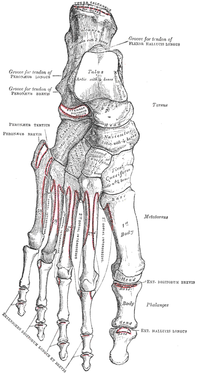
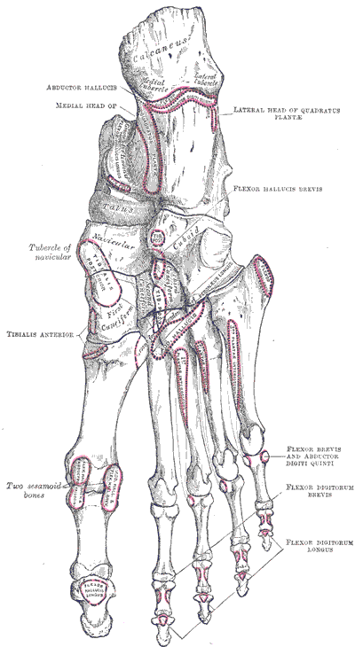
There are 7 tarsal bones, 5 metatarsal bones (the big toe is number 1, the little toe is number 5), and 14 phalanges. The big toe has only 2 phalanges, proximal and distal, while toes 2-5 each have 3, distal, middle, and proximal.
The tarsals have specific names, which you need to learn. The pictures above will help, but working with the bones in your bone box will help much more. Starting posteriorly, the bone you feel at the very back of your foot is the calcaneus, and on the top of it you find the talus. The talus articulates with the calcaneus below, the tibia and fibula above and on the sides, and the navicular bone in front. Anterior to the navicular, you find the three cuneiforms, medial, intermediate, and lateral. Finally, the cuboid bone is lateral to the cuneiforms, articulating with the 4th and 5th metatarsals anteriorly, and the calcaneus bone posteriorly. (Latin, cuneiform = wedge-shaped, calcaneum = heel, navicula = boat)
Arches:
There are two longitudinal arches, medial and lateral. The higher medial arch passes from the calcaneus to the talus to the navicular to the cuneiforms to the metatarsals. They are supported by the plantar calcaneonavicular ligament (spring ligament), and also by the tendons of the tibialis anterior and tibialis posterior muscles. The transverse arch results from the shape of the distal row of tarsal bones and the bases of the metatarsal bones. Imagine an arch that goes over the top of the cunieforms and cuboid, and is supported by the shape of the bones, various ligaments, and the tendons of the fibularis longus, tibialis anterior, and tibialis posterior muscles.
Joints:
The subtalar joint is formed between the large concave facet on the under surface of the body of the talus and the convex posterior articular surface on the superior aspect of the calcaneus. A loose, thin walled articular capsule unites the two bones by attaching to the margins of the articular surfaces. The transverse tarsal joint is the designation for the irregular plane which extends from side to side across the foot and is composed of the talonavicular articulation medially and the calcaneocuboid joint laterally. This joint allows inversion and eversion of the foot.
Ligaments:
The plantar ligaments of the joints of the foot are very strong. They they are supplemented by robust interosseous ligaments which keep the bones from spreading apart. Notable on the sole of the foot are the long plantar ligament and the plantar calcaneoucuboid and plantar calcaneonavicular ligaments. The elasticity of the latter and its support of the head of the talus have led to it being called the "spring ligament." The plantar aponeurosis may be likened to a "tie rod" for the longitudinal arch, firmly connecting its ends and preventing their spread.The dorsum of the foot is a single compartment, while the plantar foot is divided into four compartments, the medial, lateral, central, and adductor-interosseous compartments.6. Identify the muscles of the foot and give their functional significance in locomotion. (W&B 623-632, TG3-49, TG3-50, TG3-51, TG3-52A, TG3-52B)
The muscles are best considered in their different compartments. Dorsum of the foot:7. Identify the vascular supply of the foot and give the regions supplied by each. (W&B 623-632, TG3-49, TG3-50, TG3-51)
Muscle Origin Insertion Action Innervation extensor digitorum brevis superolateral surface of calcaneus extensor expansion of toes 2-4 (tendon to hallux is called extensor hallucis brevis) extends toes 2-4 deep fibular (peroneal) nerve extensor hallucis brevis superolateral surface of calcaneus dorsum of base of proximal phalanx of hallux extends great toe deep fibular (peroneal) nerve
We must also consider the extensor expansions. The three tendons of the extensor digitorum brevis join the lateral sides of the tendons of the extensor digitorum longus muscle to the second, third and fourth toes. These form the extensor expansion on these digits.
Blood supply to these muscles is via the dorsalis pedis artery, which comes from the anterior tibial artery.
Plantar Foot:
Medial Compartment:
Muscle Origin Insertion Action Innervation abductor hallucis medial side of tuberosity of calcaneus medial side of base of proximal phalanx of hallux abducts hallux; flexes metatarsophalangeal joint medial plantar nerve flexor hallucis brevis cuboid, lateral cuneiform, medial side of first metatarsal medial belly: medial side of proximal phalanx of hallux; lateral belly: lateral side of proximal phalanx flexes metatarsophalangeal joint of hallux medial plantar nerve (lateral belly occasionally receives innervation from lateral plantar nerve)
Blood supply to these muscles is via the medial plantar artery, which is from posterior tibial artery.
Lateral compartment:
Central compartment:The central compartment also technically contains the tendons of the flexor digitorum longus muscle. Also, note the split innervation of this compartment. Blood supply to the flexor digitorum brevis muscle is from both the medial and lateral plantar arteries, while the quadratus plantae muscle only receives the lateral plantar artery. Adductor-interosseous compartment:
Muscle Origin Insertion Action Innervation flexor digitorum brevis tuberosity of calcaneus, plantar aponeurosis, intermuscular septae base of middle phalanx of digits 2-5 after splitting to allow passage of flexor digitorum longus tendons flexes metatarsophalangeal & proximal interphalangeal joints of digits 2-5 medial plantar nerve quadratus plantae anterior portion of calcaneus & long plantar ligament tendons of flexor digitorum longus m. assists flexor digitorum longus in flexing toes lateral plantar nerve The adductor hallucis muscle receives blood from the plantar arterial arch, the plantar interosseous muscles get plantar metatarsal arteries, and the dorsal interosseous muscles get dorsal metatarsal arteries.
Muscle Origin Insertion Action Innervation adductor hallucis oblique head: bases of metatarsals 2-4; transverse head: heads of metatarsals 3-5 lateral side of base of proximal phalanx of hallux adducts great toe (moves it toward midline of foot; i.e. 2nd digit) deep branch of lateral plantar nerve dorsal interosseous, of foot four muscles, from shafts of adjacent metatarsal bones bases of proximal phalanges for digit 2 (both sides) & digits 3,4 (lateral side) abduct digits 2-4 (move these digits away from midline as defined by a line passing through 2nd digit), flex metatarsophalangeal joints and extend interphalangeal joints of those digits deep branch of lateral plantar nerve plantar interosseous, of foot base and medial side of metatarsals 3-5 bases of proximal phalanges and extensor expansions of digits 3-5 adduct digits 3-5 (move these digits toward the midline of the foot as defined by the second digit),flex metacarpophalangeal and extend interphalangeal joints of digits 3-5 deep branch of lateral plantar nerve
In summary, think about what you see as you take off the layers of the foot. First, you are confronted with the plantar aponeurosis. As soon as you peel it off, you see the flexor digitorum brevis muscle which runs from the calcaneus to mid-sole, and then has 4 tendons that go to toes 2-5. If you reflect it, you then see the flexor digitorum longus tendon and lateral to it is quadratus plantae muscle. (The latter muscle helps longus flex the toes in a straight line. Look at the tendon running from the medial malleous obliquely across the sole. By itself it would flex the toes medially.) Reflect this layer, and you see the adductor hallucis muscle. It is the one with both a transverse and an oblique head. You will also see the interosseous muscles between the metatarsal bones. The dorsal interosseous muscles abduct (DAB) the 2nd, 3rd, and 4th toe. (Since the midline is the 2nd toe, one can abduct this toe in two directions. Hence, it gets 2 muscles, one on either side, for abduction. The 3rd and 4th toes each get one of these muscles, and because one wants to abduct the toes, the muscles must be on the lateral side of the metatarsals. The big toe and the little toe have their own abductors.) The plantar interosseous muscles adduct (PAD) the 3rd, 4th, and 5th toes, i.e. pull them towards the second toe. (Therefore, these muscles must be on medial side of the bone. The second toe's adduction occurs simply by relaxing the abductors. The big toe has its own adductor muscle.)The dorsalis pedis, the continuation of the anterior tibial artery, has many branches, and eventually becomes the deep plantar artery. This artery dives to the sole of the foot (between the 2 heads of the 1st dorsal interosseous muscle, between the 1st and 2nd toes). It unites with the lateral plantar artery to form the plantar arterial arch.8. Identify the nerves of the foot, and the muscles and cutaneous regions supplied by them, so that given a functional and/or cutaneous loss one can predict the nerve and the probable level of injury. (W&B 581, 621-622, TG3-49, TG3-50, TG3-51)
The posterior tibial artery divides into the medial and lateral plantar arteries. The medial plantar artery runs in the groove between the medial and central compartments. It supplies the medial compartment, including the muscles of the great toe. It also gives off most of the plantar digital branches. The lateral plantar artery supplies the lateral compartment, including the muscles of the little toe. Both the lateral and medial plantar arteries supply the central compartment.
As for the innervation of muscles, the dorsum of the foot gets the deep fibular nerve. The lateral plantar compartment gets the lateral plantar nerve, the medial plantar compartment gets the medial plantar nerve, and the central compartment gets both. These plantar nerves result from the forking of the tibial nerve in the sole of the foot.
Cutaneous nerve distribution in the leg and foot: see Objective 3 above.
Cultural enrichment: Check out these sections from the 1918 version of Gray's Anatomy of the Human Body! Some of the terms are (of course) out-of-date, but the illustrations are timeless. The Muscles and Fasciae of the Leg - The Fasciae Around the Ankle - The Veins of the Lower Extremity, Abdomen, and Pelvis - Nerves - Surface Anatomy of the Lower Extremity - Surface Markings of the Lower Extremity
Questions and Answers:
9. Examine the articulation between the talus and calcaneus (the subtalar joint) and study the transverse tarsal joint, which is a complex joint that consists of the talonavicular joint and the calcaneocuboid joint. What are their functions?The transverse tarsal joint refers to an irregular articular plane which extends from side to side across the foot and is made of the talonavicular articulation medially and the calcaneocuboid joints laterally. At this plane primarily, and at other tarsal joints to a lesser degree, are produced the movements of inversion and eversion of the foot. With inversion is the combined adduction and plantar flexion; with eversion, abduction and dorsiflexion. The contribution of the subtalar and talocalcaneonavicular joints is that of movement around the axis that passes through the tarsal sinus. These movements allow the foot to be placed firmly on slanting and irregular surfaces and still serve as a firm basis of support for the body. (TG3-61)10. Does the anterior lateral malleolar artery communicate with the perforating artery?Yes. The anterior lateral malleolar artery comes off the anterior tibial artery around the ankle; it goes to the lateral malleolus. The fibular (peroneal) artery, off the posterior tibial artery, gives off a perforating branch that passes forward at the distal border of the interosseous membrane and anastomoses with the anterior lateral malleolar artery. (TG3-38)11. Trace the deep fibular (peroneal) nerve into the anterior compartment of the leg with its accompanying artery and vein. Note how it innervates anterior compartment muscles and continues into the foot. What muscle does it supply there? To what area does it supply cutaneous Innervation?The deep fibular nerve serves the tibialis anterior muscle, the extensor digitorum longus muscle, the extensor hallucis longus muscle, and the fibularis (peroneus) tertius muscle. It also provides articular branches to the tibiofibular syndesmosis and the ankle joint. After crossing the ankle, the deep fibular nerve divides into medial and lateral branches, which supply the dorsum of the foot, and supplies the extensor digitorum brevis and extensor hallucis brevis muscles. The medial branch divides into two dorsal digit branches which supply adjacent sides of the first and second digits. Twigs also go to the metatarsophalangeal and interphalangeal articulations of the great toe and one to the first dorsal interosseous muscle. The lateral branch passes laterally, deep to the extensor digitorum brevis muscle. It ends in an enlargement from which branches distribute to this muscle, the tarsal joints, and the three lateral intermetatarsal spaces for the supply of the periosteum and the joints. The cutaneous innervation supplied by this nerve is on the dorsum of the foot, between the second and big toe. (TG3-37)12. Which nerve supplies the muscles of the lateral compartment?The lateral compartment has two muscles: fibularis (peroneus) longus and fibularis (peroneus) brevis. Innervation is via the superficial fibular (peroneal) nerve. (TG3-36)13. What innervates the flexor digitorum brevis muscle?The medial plantar nerve, from the tibial nerve, innervates the flexor digitorum brevis muscle. (TG3-49)14. Define the axis of adduction and abduction in the foot and observe how each of the plantar and dorsal interosseous muscles fits into the plan.The axis of abduction or adduction of digits of the foot is a line through the second toe. (Note that, in the hand, the axis runs through the third finger.)15. What is the plantar arterial arch?
See #7 above.The lateral plantar artery, off of the posterior tibial artery, crosses the sole of the foot diagonally from the medial to the lateral side. At the medial side of the base of the 5th metatarsal bone, the artery turns medially around the margin of the quadratus plantae muscle and sinks between the adductor hallucis and interosseous muscles. It perforates the plantar interosseous fascia and passes medially across the proximal ends of the second, third, and fourth metatarsal bones and the corresponding interosseous muscles. Here it forms the plantar arterial arch, anastamosing in the first interosseous space with the deep plantar branch of the dorsalis pedis artery. (TG3-51)